. 2019 May 1;5(5):eaaw1616.
doi: 10.1126/sciadv.aaw1616. eCollection 2019 May.

Structural mechanism for regulation of the AAA-ATPases RUVBL1-RUVBL2 in the R2TP co-chaperone revealed by cryo-EM

Affiliations

Structural mechanism for regulation of the AAA-ATPases RUVBL1-RUVBL2 in the R2TP co-chaperone revealed by cryo-EM

Hugo Muñoz-Hernández et al. Sci Adv. .

Abstract

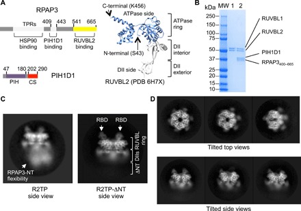
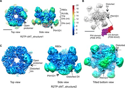
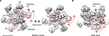
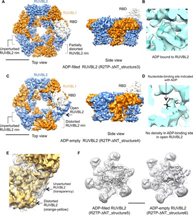
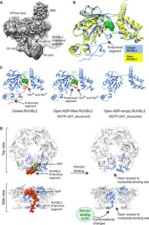
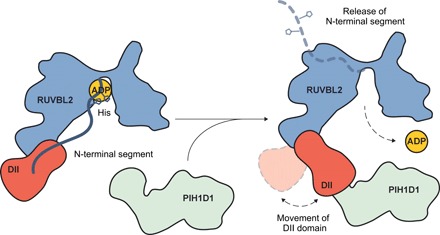
The human R2TP complex (RUVBL1-RUVBL2-RPAP3-PIH1D1) is an HSP90 co-chaperone required for the maturation of several essential multiprotein complexes, including RNA polymerase II, small nucleolar ribonucleoproteins, and PIKK complexes such as mTORC1 and ATR-ATRIP. RUVBL1-RUVBL2 AAA-ATPases are also primary components of other essential complexes such as INO80 and Tip60 remodelers. Despite recent efforts, the molecular mechanisms regulating RUVBL1-RUVBL2 in these complexes remain elusive. Here, we report cryo-EM structures of R2TP and show how access to the nucleotide-binding site of RUVBL2 is coupled to binding of the client recruitment component of R2TP (PIH1D1) to its DII domain. This interaction induces conformational rearrangements that lead to the destabilization of an N-terminal segment of RUVBL2 that acts as a gatekeeper to nucleotide exchange. This mechanism couples protein-induced motions of the DII domains with accessibility of the nucleotide-binding site in RUVBL1-RUVBL2, and it is likely a general mechanism shared with other RUVBL1-RUVBL2-containing complexes.


Figures

Fig. 1
Fig. 1. Purification and cryo-EM of R2TP-ΔNT.
(A) Left: Components of R2TP and their domains. TPR, PIH, and CS domains are indicated. Right: Domains of a RUVBL2 subunit (PDB 6H7X, residues S43 to K456) (26), indicating the ATPase and DII side of the ring. (B) SDS-polyacrylamide gel electrophoresis for the final purification steps of RUVBL1-RUVBL2 (lane 1), which we mixed with the RPAP3400–665-PIH1D1 (lane 2) subcomplex. We used the sample in lane 2 for vitrification and cryo-EM. MW, molecular weight markers. (C) Representative side view 2D average of R2TP (17) and the truncated version produced in this work. Flexible regions in R2TP disappear in R2TP-ΔNT after the first 399 residues of RPAP3 are removed. RBD domains are labeled with white arrows, and the location of the RUVBL ring and DII domains is indicated. Images are at a different scale. (D) In the micrographs, multiple orientations of top, tilted, and side views were found, assuring a good coverage of Euler angles during image processing.
Fig. 2
Fig. 2. Overall architecture of R2TP-ΔNT.
(A) Consensus 3D structure of R2TP-ΔNT at 4.0 Å resolution (R2TP-ΔNT_structure1) colored according to local resolution (40). The structure shows an opening in one end, as well as the location of PIH1D1 at the DII face of the ring. Color code scale is shown. (B) Classification of the particles based on the variability of PIH1D1 into three subgroups. The most abundant group shows clear density for PIH1D1 and the displaced DII domain, and we selected this for further refinement. A view of the DII face of the ring reveals density for PIH1D1, which agrees in shape and size with crystal structures of the PIH and CS domains, colored purple and red, respectively. Scale bar, 5 nm. (C) Structure of R2TP-ΔNT_structure2 obtained after refinement of the subgroup shown in (B). We estimated local resolutions in RELION (33), and we filtered and colored the map accordingly, using the same color code used in (A). PIH1D1, the DII domain interacting with PIH1D1, and the distorted rim of the ring are indicated. The top view is shown with a slightly higher threshold than the side and tilted views, to highlight the opening of one protomer and the distorted rim.
Fig. 3
Fig. 3. Motions of PIH1D1.
(A) After principal components analysis, we generated 10 maps, each representing the median orientation of the particles within each specific subset. A bottom view of two of these maps is shown, highlighting the movement of PIH1D1 within the structure. The mobile element, comprising PIH1D1 and the DII domain, is enclosed within a red line. (B) Side view of one of the maps generated after multibody refinement. Observed motions are proximal to regions of the RUVBL1-RUVBL2 ring, suggesting that conformational changes in the DII domain could be coupled to changes in the ring. A red line is used to highlight the proximity between the mobile elements and the RUVBL1-RUVBL2 ring.
Fig. 4
Fig. 4. ADP-filled and ADP-empty conformations of the open RUVBL2 subunit.
(A) Top and side view of the 3D structure of the ADP-filled open conformation of the RUVBL1-RUVBL2 ring bound to one RBD domain (R2TP-ΔNT_structure3). Color code: orange, RUVBL1; blue, RUVBL2; and white, RBD. Scale bar, 5 nm. (B) Close-up of the nucleotide-binding site of the open RUVBL2 subunit in R2TP-ΔNT_structure3, showing density for ADP, represented in black. (C) Top view and side view of the 3D structure of the ADP-empty open conformation of the RUVBL1-RUVBL2 ring bound to one RBD domain (R2TP-ΔNT_structure4). Color code: orange, RUVBL1; blue, RUVBL2; and white, RBD. Scale bar, 5 nm. (D) Close-up of the nucleotide-binding site of the open RUVBL2 subunit in R2TP-ΔNT_structure4, showing the absence of density in the nucleotide-binding site. ADP in the putative location of the nucleotide-binding site is represented in black to highlight the absence of density. (E) Close-up comparison between the rim of an unperturbed (transparent density) and a distorted RUVBL2 subunit (orange-yellow) from R2TP-ΔNT_structure5 represented at identical threshold. (F) Views of the DII face of the ring of the R2TP-ΔNT_structure5 and R2TP-ΔNT_structure6 structures, obtained from the particles classified as ADP-filled and ADP-empty but without subtracting the PIH1D1 region. The location of PIH1D1 in the structure is indicated within a dashed black line. Scale bar, 5 nm.
Fig. 5
Fig. 5. Mechanism of RUVBL1-RUVBL2 remodeling and nucleotide exchange.
(A) A view of the map showing density for the N-terminal loop in RUVBL2 contacting the DII domain of the same protomer. (B) The structures of RUVBL2 closed (blue) and ADP-empty conformation (yellow) are superimposed. For alignment of the two structures, we used the neighboring RUVBL1 subunit as template. Thus, the changes observed represent movements in the context of the RUVBL ring. ADP is shown in green. (C) Structures of the RUVBL2 subunit in closed, open ADP-filled, and open ADP-empty conformation. ADP is shown in green. The N-terminal segment (residues 8 to 49 in the closed structure and residues 23 to 49 in the open ADP-filled structure) is colored red, indicating the position of His25 and His27. (D) Side view and top view of the closed and open structure of the RUVBL1-RUVBL2 hexamer, shown in ribbons, and highlighting one RUVBL2 subunit in blue. The RUVBL2 N-terminal segment shown as spheres and in red blocks the access to the nucleotide-binding site, and this obstruction is removed after PIH1D1 binds the DII domain and induces conformational changes.
Fig. 6
Fig. 6. Model for remodeling of RUVBL1-RUVBL2 by PIH1D1.
Cartoon for the mechanistic model of how PIH1D1 regulates the accessibility to the nucleotide-binding site of RUVBL1-RUVBL2.

References

    1. Machado-Pinilla R., Liger D., Leulliot N., Meier U. T., Mechanism of the AAA+ ATPases pontin and reptin in the biogenesis of H/ACA RNPs. RNA 18, 1833–1845 (2012). - PMC - PubMed
    1. Horejsi Z., Takai H., Adelman C. A., Collis S. J., Flynn H., Maslen S., Skehel J. M., de Lange T., Boulton S. J., CK2 phospho-dependent binding of R2TP complex to TEL2 is essential for mTOR and SMG1 stability. Mol. Cell 39, 839–850 (2010). - PubMed
    1. Cloutier P., Poitras C., Durand M., Hekmat O., Fiola-Masson É., Bouchard A., Faubert D., Chabot B., Coulombe B., R2TP/Prefoldin-like component RUVBL1/RUVBL2 directly interacts with ZNHIT2 to regulate assembly of U5 small nuclear ribonucleoprotein. Nat. Commun. 8, 15615 (2017). - PMC - PubMed
    1. Kim S. G., Hoffman G. R., Poulogiannis G., Buel G. R., Jang Y. J., Lee K. W., Kim B. Y., Erikson R. L., Cantley L. C., Choo A. Y., Blenis J., Metabolic stress controls mTORC1 lysosomal localization and dimerization by regulating the TTT-RUVBL1/2 complex. Mol. Cell 49, 172–185 (2013). - PMC - PubMed
    1. Takai H., Xie Y., de Lange T., Pavletich N. P., Tel2 structure and function in the Hsp90-dependent maturation of mTOR and ATR complexes. Genes Dev. 24, 2019–2030 (2010). - PMC - PubMed

Publication types

MeSH terms