Review
. 2019 Sep;76(18):3479-3496.
doi: 10.1007/s00018-019-03104-6. Epub 2019 May 3.

Human placenta and trophoblast development: key molecular mechanisms and model systems

Affiliations
Review

Human placenta and trophoblast development: key molecular mechanisms and model systems

Martin Knöfler et al. Cell Mol Life Sci. 2019 Sep.

Abstract

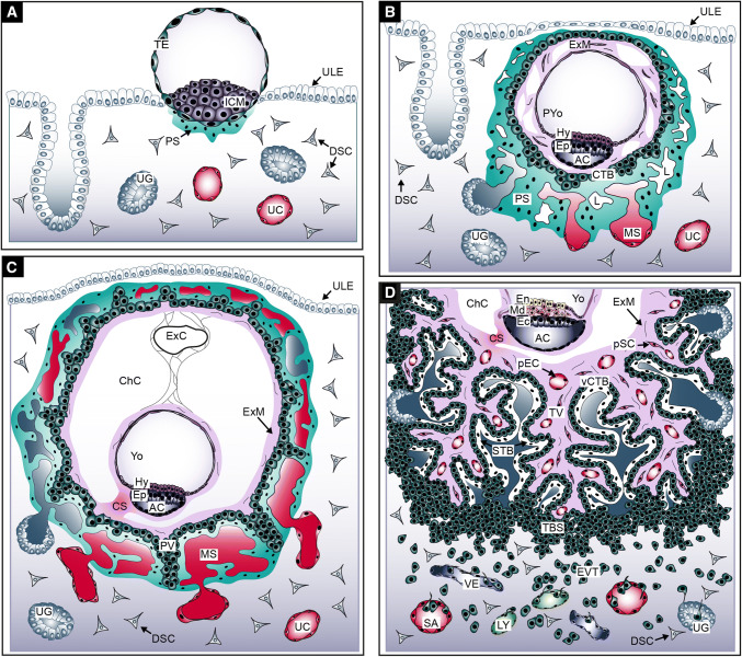
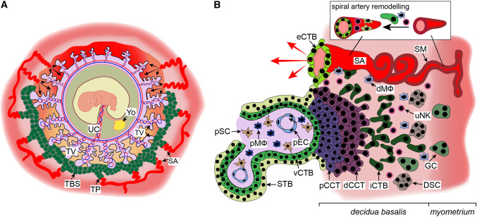
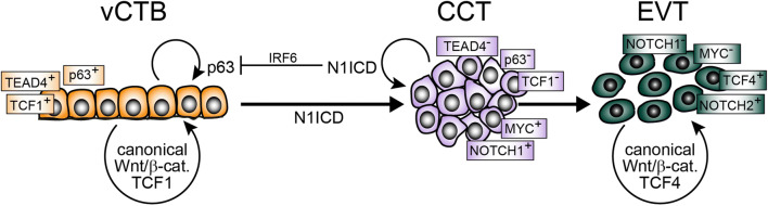
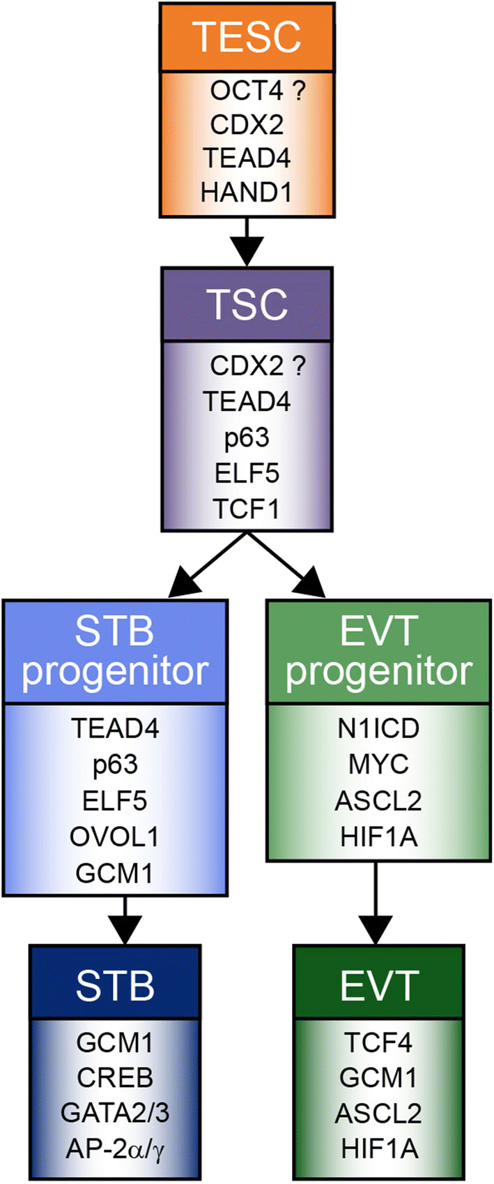
Abnormal placentation is considered as an underlying cause of various pregnancy complications such as miscarriage, preeclampsia and intrauterine growth restriction, the latter increasing the risk for the development of severe disorders in later life such as cardiovascular disease and type 2 diabetes. Despite their importance, the molecular mechanisms governing human placental formation and trophoblast cell lineage specification and differentiation have been poorly unravelled, mostly due to the lack of appropriate cellular model systems. However, over the past few years major progress has been made by establishing self-renewing human trophoblast stem cells and 3-dimensional organoids from human blastocysts and early placental tissues opening the path for detailed molecular investigations. Herein, we summarize the present knowledge about human placental development, its stem cells, progenitors and differentiated cell types in the trophoblast epithelium and the villous core. Anatomy of the early placenta, current model systems, and critical key regulatory factors and signalling cascades governing placentation will be elucidated. In this context, we will discuss the role of the developmental pathways Wingless and Notch, controlling trophoblast stemness/differentiation and formation of invasive trophoblast progenitors, respectively.

Keywords: Chorionic villus; Mesenchymal cell; Placenta development; Trophoblast differentiation; Trophoblast stem cell.


Figures

Fig. 1
Fig. 1
Development of the human placenta during the first 3 weeks of gestation. a Human blastocyst implanting into the pregnant uterus. b Development of the first placental structures and the embryonic disc. c Formation of primary villi and yolk sac. d Development of tertiary villi and the embryonic germ layers. AC amniotic cavity, CS connecting stalk, ChC chorionic cavity, CTB cytotrophoblast, DSC decidual stromal cell, Ec ectoderm, En endoderm, Ep epiblast, EVT extravillous trophoblast, ExC exocoelomic cyst, ExM extraembryonic mesoderm, Hy hypoblast, ICM inner cell mass, L lacunae system, LY lymphatic vessel, Md mesoderm, MS maternal blood sinusoid, pEC placental endothelial cell, PS primitive syncytium, pSC placental stromal cell, PV primary villi, PYo primitive yolk sac, SA spiral artery, TBS trophoblastic shell, TV tertiary villi, UC uterine capillary, UG uterine gland, ULE uterine luminal epithelium, VE venous vessel, vCTB villous CTB, Yo yolk sac
Fig. 2
Fig. 2
Development of the trophoblastic shell and formation of placental anchoring villi. a Structure of the human trophoblastic shell and its surrounding arterial vessels. b Depiction of a placental anchoring villus, spiral artery (SA) remodelling and interaction of extravillous trophoblasts (EVTs) with different decidual cell types. dCCT distal cell column trophoblast, dMΦ decidual macrophage, DSC decidual stromal cell, eCTB endovascular cytotrophoblast, GC giant cell, iCTB interstitial cytotrophoblast, pCCT proximal cell column trophoblast, pEC placental endothelial cell, pMΦ placental macrophage, pSC placental stromal/mesenchymal cell, SM smooth muscle layer, STB syncytiotrophoblast, TBS trophoblastic shell, TP trophoblast plug, TV tertiary villi, UC umbilical cord, uNK uterine NK cell, vCTB villous cytotrophoblast, YO yolk sac
Fig. 3
Fig. 3
Model system integrating the role of Notch/Wnt signalling in trophoblast stemness and EVT differentiation. Notch1 intracellular domain (N1ICD) represses markers of villous cytotrophoblast (vCTB) self-renewal, i.e. TEAD4 and p63, and induces expression of the extravillous trophoblast (EVT)-progenitor-specific gene MYC. Formation of these precursors is also associated with the loss of TCF1 expression, whereas β-catenin–TCF4 complexes arise during EVT formation. CCT cell column trophoblast, IRF6 interferon regulatory factor 6, TCF T-cell factor, Wnt wingless
Fig. 4
Fig. 4
Trophoblast lineage development and key regulatory transcription factors expressed in the different trophoblast subtypes. Regulators, operating in both syncytiotrophoblast (STB) and extravillous trophoblast (EVT), are not depicted. Likewise, distal cell column trophoblasts, developing from EVT progenitors are not shown, since these cells express the same repertoire of key transcription factors as EVTs. Further differentiation steps of these cells in deeper regions of the decidua were omitted, since knowledge about the associated transcription factors is lacking. AP-2α, AP-2γ and GATA3 represent reliable markers of trophoblast identity, yet are present in most trophoblast subtypes. Herein, presentation of these factors only in STB aims indicating their predominant roles in placental hormone expression. A possible role of OCT4 in the pre-implantation trophectodermal stem cell (TESC) remains disputable. The absence of CDX2 from the majority of CTBs of self-renewing cultures, questions its role in post-implantation trophoblast stem cell (TSC) maintenance. TSCs are potentially equivalent to self-renewing villous cytotrophoblasts of the placental epithelium

Comment in

Similar articles

Cited by

References

    1. Hamilton WJ, Boyd JD. Development of the human placenta in the first 3 months of gestation. J Anat. 1960;94:297–328. - PMC - PubMed
    1. Burton GJ, Fowden AL. The placenta: a multifaceted, transient organ. Philos Trans R Soc Lond B Biol Sci. 2015;370:20140066. doi: 10.1098/rstb.2014.0066. - DOI - PMC - PubMed
    1. Burton GJ, Charnock-Jones DS, Jauniaux E. Regulation of vascular growth and function in the human placenta. Reproduction. 2009;138:895–902. doi: 10.1530/REP-09-0092. - DOI - PubMed
    1. Napso T, Yong HEJ, Lopez-Tello J, Sferruzzi-Perri AN. The role of placental hormones in mediating maternal adaptations to support pregnancy and lactation. Front Physiol. 2018;9:1091. doi: 10.3389/fphys.2018.01091. - DOI - PMC - PubMed
    1. Mastorakos G, Ilias I. Maternal and fetal hypothalamic-pituitary-adrenal axes during pregnancy and postpartum. Ann N Y Acad Sci. 2003;997:136–149. doi: 10.1196/annals.1290.016. - DOI - PubMed

LinkOut - more resources