Review
. 2019 Nov;44(11):3518-3526.
doi: 10.1007/s00261-019-02037-9.

Complete mesocolic excision and central vascular ligation for right colon cancer: an introduction for abdominal radiologists

Affiliations
Review

Complete mesocolic excision and central vascular ligation for right colon cancer: an introduction for abdominal radiologists

David D B Bates et al. Abdom Radiol (NY). 2019 Nov.

Abstract

Objective: To provide an overview of complete mesocolic excision, along with a review of the relevant vascular anatomy and locoregional staging concepts, for abdominal radiologists.

Results: Complete mesocolic excision (CME) with central vascular ligation (CVL) for colon cancer has emerged as a technique that has growing interest in surgical oncology. Specific anatomic considerations and patterns of nodal spread have thus gained clinical significance, and should be familiar to abdominal radiologists. This review article provides an overview of CME with CVL, and discusses some of the important anatomic considerations in patients with colon cancer that are relevant to radiologists.

Conclusion: Knowledge of CME with CVL and the relevant anatomic and staging considerations is important for abdominal radiologists, as this surgical technique becomes increasingly utilized.

Keywords: Central vascular ligation; Colon cancer; Colorectal cancer; Complete mesocolic excision.


Figures
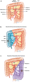
Figure 1:
Figure 1:
(a) Overview of standard arterial anatomy of the colon. The superior mesenteric artery (SMA) gives rise to the ileocolic artery (ICA), right colic artery (RCA), and middle colic artery (MCA). The inferior mesenteric artery (23) gives rise to the left colic artery (LCA), supplying the left colon from the splenic flexure to the upper rectum. The right and left-sided arterial supply are integrated by the marginal artery of Drummond. (b) Diagram showing standard vascular supply to the right colon, with an ascending colonic mass. (c) Diagram showing standard vascular supply to the left colon, with a descending colonic mass.
Figure 2:
Figure 2:
Diagram showing variant anatomy and antero-posterior relationship of rightsided arteries and veins. (a) The right colic artery (RCA) is present (60.1% of cases) and courses anterior to the superior mesenteric vein (SMV) (89.4% of the time). (b) The RCA is absent and the ileocolic artery (ICA) is anterior to the ileocolic vein (ICV) (42.6% of the time). (c) The ICA is shown in its more common relationship, posterior to the ICV (57.4% of cases).
Figure 3:
Figure 3:
Right colic artery coursing posterior to the superior mesenteric vein. Axial, sagittal and coronal contrast enhanced CT images show the RCA (arrows) positioned posterior to the SMV.
Figure 4:
Figure 4:
Right colic artery coursing anterior to the superior mesenteric vein. Axial, sagittal and coronal contrast enhanced CT images show the RCA (arrows) positioned anterior to the SMV.
Figure 5:
Figure 5:
Ileocolic artery coursing posterior to the ileocolic vein. Axial, sagittal and coronal contrast enhanced CT images show the ICA (arrows) posterior to the SMV.
Figure 6:
Figure 6:
Ileocolic artery coursing anterior to the ileocolic vein. Axial, sagittal and coronal contrast enhanced CT images show the ICA (arrows) anterior to the SMV.
Figure 7:
Figure 7:
Axial and coronal contrast enhanced CT images show a large mass (a, arrow) in the ascending colon, with an enlarged ileocolic lymph node (b and c, arrows).
Figure 8:
Figure 8:
Axial, coronal and sagittal contrast enhanced CT images show a mass in the hepatic flexure (arrowheads) with adenopathy in the root of the mesentery (arrows), as well as hepatic metastases (asterisk).
Figure 9:
Figure 9:
Coronal and sagittal contrast enhanced CT images show a mass in the hepatic flexure/proximal transverse colon (a and b, arrowheads) with cluster of small mesocolic nodes (b, arrows) and moderately suspicious gastrocolic adenopathy (c, arrow).
Figure 10:
Figure 10:
Multiplanar reformatted image from a contrast enhanced CT showing the gastrocolic trunk of Henle (GTH). A: Superior mesenteric vein; B: Middle colic vein; C: Gastrocolic trunk; D: Right Gastroepiploic vein; E: Anterosuperior pancreaticoduodenal vein; F: Superior right colic; G: Right colic vein.

References

    1. Heald RJ, Husband EM, Ryall RD. The mesorectum in rectal cancer surgery--the clue to pelvic recurrence? Br J Surg. 1982;69(10):613–6. - PubMed
    1. Maurer CA, Renzulli P, Kull C, Kaser SA, Mazzucchelli L, Ulrich A, et al. The impact of the introduction of total mesorectal excision on local recurrence rate and survival in rectal cancer: long-term results. Ann Surg Oncol. 2011;18(7):1899–906. - PubMed
    1. Beets-Tan RGH, Lambregts DMJ, Maas M, Bipat S, Barbaro B, Curvo-Semedo L, et al. Magnetic resonance imaging for clinical management of rectal cancer: Updated recommendations from the 2016 European Society of Gastrointestinal and Abdominal Radiology (ESGAR) consensus meeting. European radiology. 2018;28(4):1465–75. - PMC - PubMed
    1. Gollub MJ, Arya S, Beets-Tan RG, dePrisco G, Gonen M, Jhaveri K, et al. Use of magnetic resonance imaging in rectal cancer patients: Society of Abdominal Radiology (SAR) rectal cancer disease-focused panel (DFP) recommendations 2017. Abdominal radiology (New York). 2018;43(11):2893–902. - PubMed
    1. Hohenberger W, Weber K, Matzel K, Papadopoulos T, Merkel S. Standardized surgery for colonic cancer: complete mesocolic excision and central ligation--technical notes and outcome. Colorectal Dis. 2009;11(4):354–64; discussion 64-5. - PubMed

LinkOut - more resources