

A micromechanical muscle model for determining the impact of motor unit fiber clustering on force transmission in aging skeletal muscle
- PMID: 31049781
- PMCID: PMC6748884
- DOI: 10.1007/s10237-019-01152-2
A micromechanical muscle model for determining the impact of motor unit fiber clustering on force transmission in aging skeletal muscle
Abstract
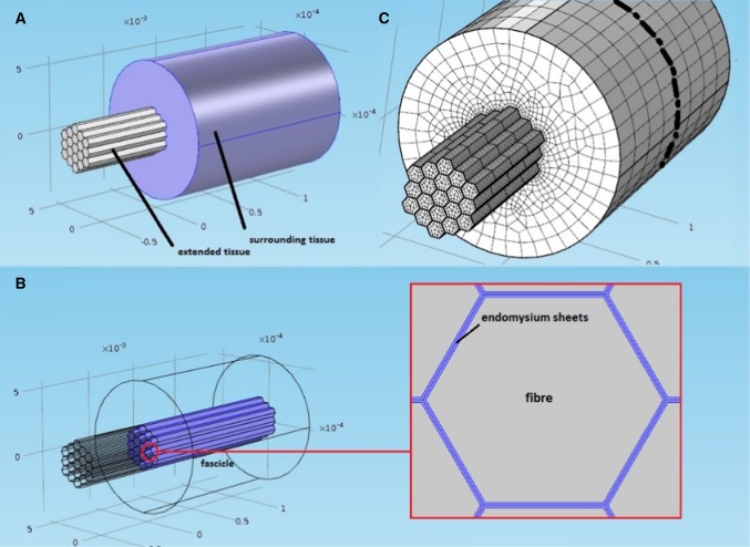
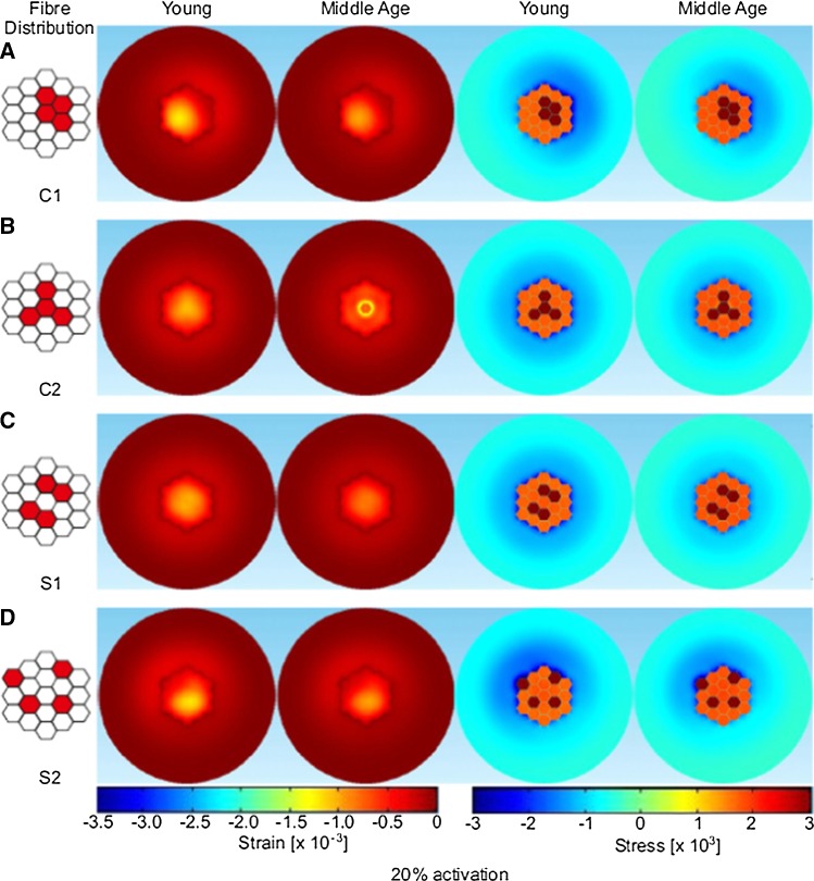
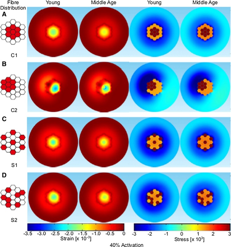
This study used a micromechanical finite element muscle model to investigate the effects of the redistribution of spatial activation patterns in young and old muscle. The geometry consisted of a bundle of 19 active muscle fibers encased in endomysium sheets, surrounded by passive tissue to model a fascicle. Force was induced by activating combinations of the 19 active muscle fibers. The spacial clustering of muscle fibers modeled in this study showed unbalanced strains suggesting tissue damage at higher strain levels may occur during higher levels of activation and/or during dynamic conditions. These patterns of motor unit remodeling are one of the consequences of motor unit loss and reinnervation associated with aging. The results did not reveal evident quantitative changes in force transmission between old and young adults, but the patterns of stress and strain distribution were affected, suggesting an uneven distribution of the forces may occur within the fascicle that could provide a mechanism for muscle injury in older muscle.
Keywords: Fiber clustering; Finite element modeling; Force; Motor unit; Muscle; Spacial activation patterns.
Conflict of interest statement
The authors declare that they have no conflict of interest.
Figures
References
-
- Ansved T, Wallner P, Larsson L. Spatial distribution of motor unit fibres in fast- and slow-twitch rat muscles with special reference to age. Acta Physiol Scand. 1991;143(3):345–354. - PubMed
-
- Ballak SB, Degens H, de Haan A, Jaspers RT. Aging related changes in determinants of muscle force generating capacity: a comparison of muscle aging in men and male rodents. Ageing Res Rev. 2014;14:43–55. - PubMed
-
- Blemker SS, Pinsky PM, Delp SL. A 3D model of muscle reveals the causes of nonuniform strains in the biceps brachii. J Biomech. 2005;38(4):657–665. - PubMed
-
- Bloch RJ, Gonzalez-Serratos H. Lateral force transmission across costameres in skeletal muscle. Exerc Sport Sci Rev. 2003;31(2):73–78. - PubMed
-
- Bosboom EM, Hesselink MK, Oomens CW, Bouten CV, Drost MR, Baaijens FP. Passive transverse mechanical properties of skeletal muscle under in vivo compression. J Biomech. 2001;34(10):1365–1368. - PubMed
MeSH terms
Grants and funding
LinkOut - more resources
Full Text Sources
Medical
