Review
. 2019 Jun;11(3):287-302.
doi: 10.1007/s12551-019-00526-1. Epub 2019 May 2.

Microbial functional amyloids serve diverse purposes for structure, adhesion and defence

Affiliations
Review

Microbial functional amyloids serve diverse purposes for structure, adhesion and defence

Nirukshan Shanmugam et al. Biophys Rev. 2019 Jun.

Abstract

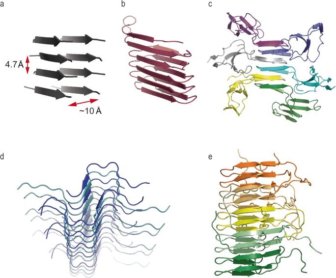
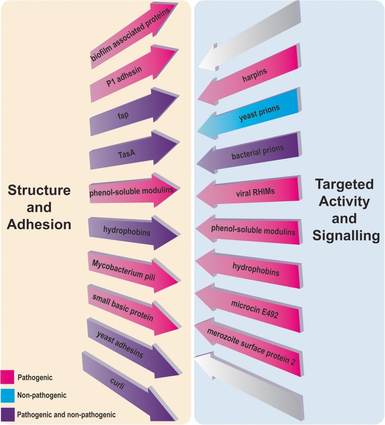
The functional amyloid state of proteins has in recent years garnered much attention for its role in serving crucial and diverse biological roles. Amyloid is a protein fold characterised by fibrillar morphology, binding of the amyloid-specific dyes Thioflavin T and Congo Red, insolubility and underlying cross-β structure. Amyloids were initially characterised as an aberrant protein fold associated with mammalian disease. However, in the last two decades, functional amyloids have been described in almost all biological systems, from viruses, to bacteria and archaea, to humans. Understanding the structure and role of these amyloids elucidates novel and potentially ancient mechanisms of protein function throughout nature. Many of these microbial functional amyloids are utilised by pathogens for invasion and maintenance of infection. As such, they offer novel avenues for therapies. This review examines the structure and mechanism of known microbial functional amyloids, with a particular focus on the pathogenicity conferred by the production of these structures and the strategies utilised by microbes to interfere with host amyloid structures. The biological importance of microbial amyloid assemblies is highlighted by their ubiquity and diverse functionality.

Keywords: Biofilm; Curli; Fibrils; Functional amyloid; Hydrophobin; RHIM.


Conflict of interest statement

Nirukshan Shanmugam declares that he has no conflict of interest. Max O. D. G. Baker declares that he has no conflict of interest. Sarah R. Ball declares that she has no conflict of interest. Megan Steain declares that she has no conflict of interest. Chi L. L. Pham declares that she has no conflict of interest. Margaret Sunde declares that she has no conflict of interest.

Figures

Fig. 1
Fig. 1
Comparison of different amyloid fibril structures. Although all amyloids have a common cross-β structure, they have different backbone conformations dependent on backbone and inter-sheet interactions. It is likely that these different conformers of the amyloid fold confer some differences in functionality. a Generic fibril structure. All amyloid structures are defined by an underlying cross-β structure, wherein two β-sheets are parallel to the fibril axis, and component β-strands lie perpendicular to the same axis. The inter-sheet distance is approximately 10 Å and the inter-strand distance is approximately 4.7 Å. b Model of an E. coli curli fibril. Curli fibrils appear on the surface of certain bacteria and provide adherence properties and structural stability. Curli fibrils are comprised of repeat monomers of the protein CsgA. CsgA is nucleated on the cell surface by accessory proteins. Left-handed β-helix model reproduced with kind permission from K. Lindorff-Larsen, adapted from Tian et al. (2015). c Model of an EASΔ15 hydrophobin rodlet from Neurospora crassa. Hydrophobins undergo a conformational change at air:water interfaces and self-associate into an amyloid-like structure termed a rodlet. Five hydrophobin monomers shown in different colours. EASΔ15 rodlet model reproduced with kind permission from A. Kwan, adapted from Macindoe et al. (2012). d Model structure of a heteromeric amyloid fibril containing M45 from murine cytomegalovirus and human RIPK3. It is hypothesised that the human and viral proteins are integrated into the same fibril by RHIM:RHIM interactions, forming a serpentine fold. The formation of this hybrid amyloid inhibits the signalling capabilities of the host protein by unknown mechanisms. Image produced using a model of RIPK3:M45 reported in (Pham et al. 2019) and based on the RIPK1:RIPK3 hetero-amyloid structure PDB 5V7V (Mompean et al. 2018). e Structure of the Het-s prion amyloid from Podospora anserina. When assembled into an amyloid structure, the Het-s protein adopts a β-solenoid conformation. This amyloid induces programmed cell death upon encountering heterokaryon incompatibility. Five protein monomers shown in different colours. Imaged produced from PDB 2RNM Wasmer et al. (2008)
Fig. 2
Fig. 2
Classification of functional amyloids utilised by bacteria, fungi, protozoa and viruses, according to their roles

References

    1. Adda CG, et al. Plasmodium falciparum merozoite surface protein 2 is unstructured and forms amyloid-like fibrils. Mol Biochem Parasitol. 2009;166:159–171. doi: 10.1016/j.molbiopara.2009.03.012. - DOI - PMC - PubMed
    1. Aimanianda V, et al. Surface hydrophobin prevents immune recognition of airborne fungal spores. Nature. 2009;460:1117–1121. doi: 10.1038/nature08264. - DOI - PubMed
    1. Alsteens D, Ramsook CB, Lipke PN, Dufrene YF. Unzipping a functional microbial amyloid. ACS Nano. 2012;6:7703–7711. doi: 10.1021/nn3025699. - DOI - PMC - PubMed
    1. Alteri CJ, Xicohtencatl-Cortes J, Hess S, Caballero-Olin G, Giron JA, Friedman RL. Mycobacterium tuberculosis produces pili during human infection. Proc Natl Acad Sci U S A. 2007;104:5145–5150. doi: 10.1073/pnas.0602304104. - DOI - PMC - PubMed
    1. Baker M, Shanmugam N, Pham CLL, Strange M, Steain M, Sunde M (2018) RHIM-based protein:protein interactions in microbial defence against programmed cell death by necroptosis. Semin Cell Dev Biol. 10.1016/j.semcdb.2018.05.004 - PubMed