

Immunocytochemistry for predictive biomarker testing in lung cancer cytology
- PMID: 31050216
- PMCID: PMC7493418
- DOI: 10.1002/cncy.22137
Immunocytochemistry for predictive biomarker testing in lung cancer cytology
Abstract
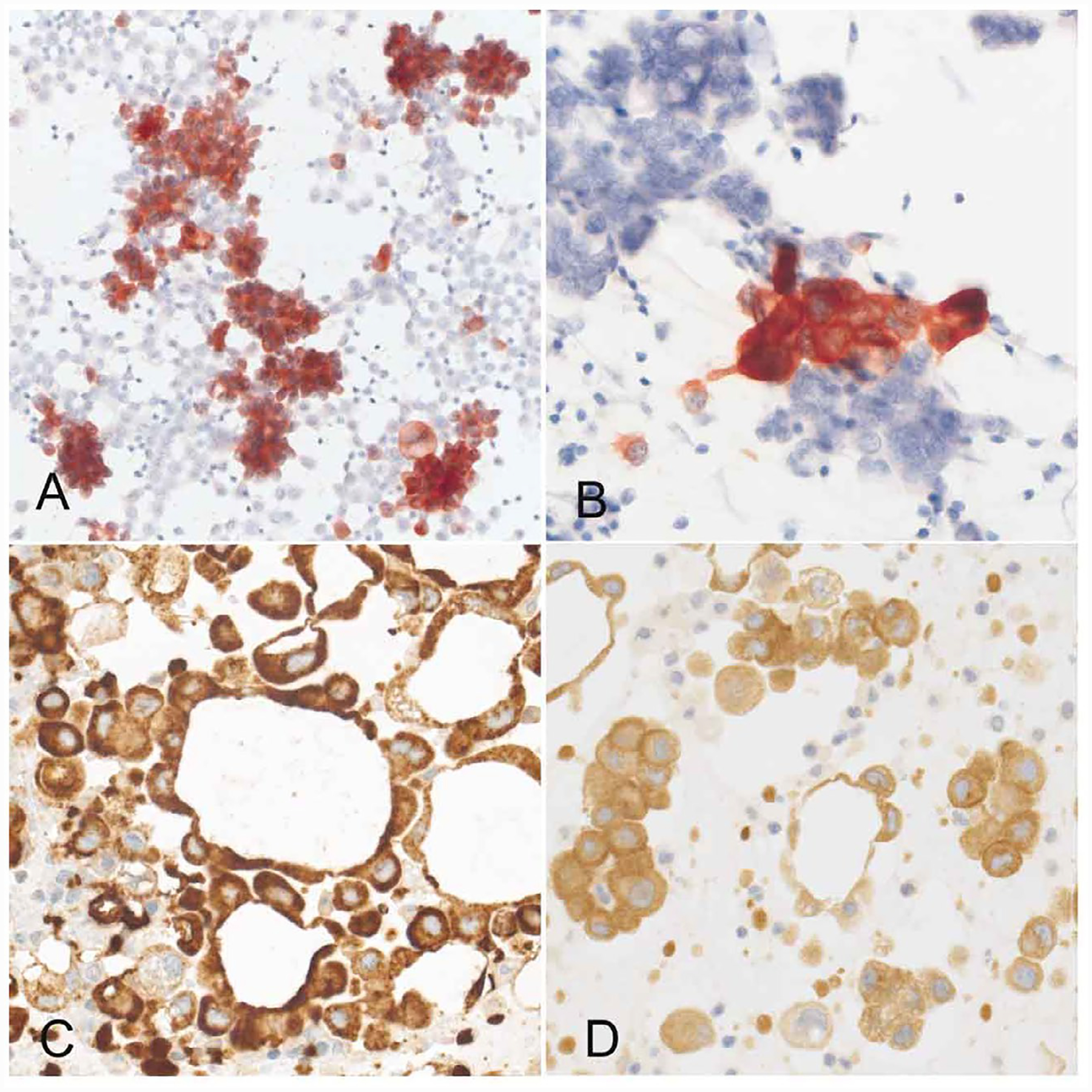
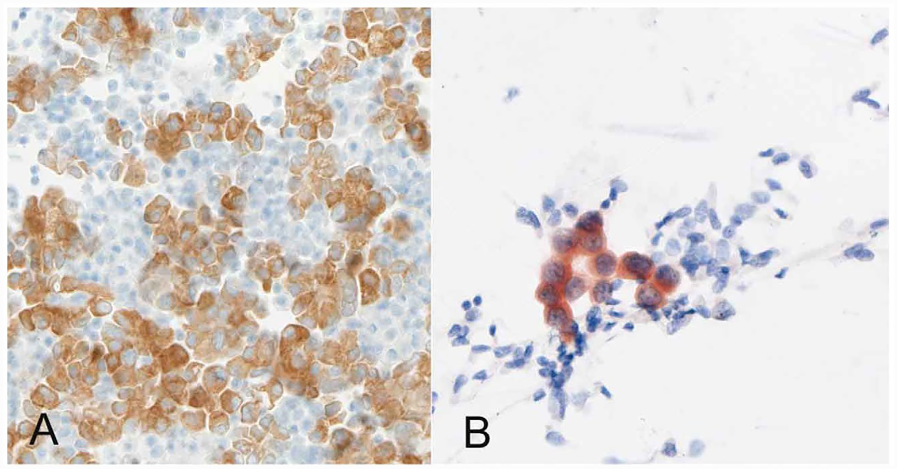
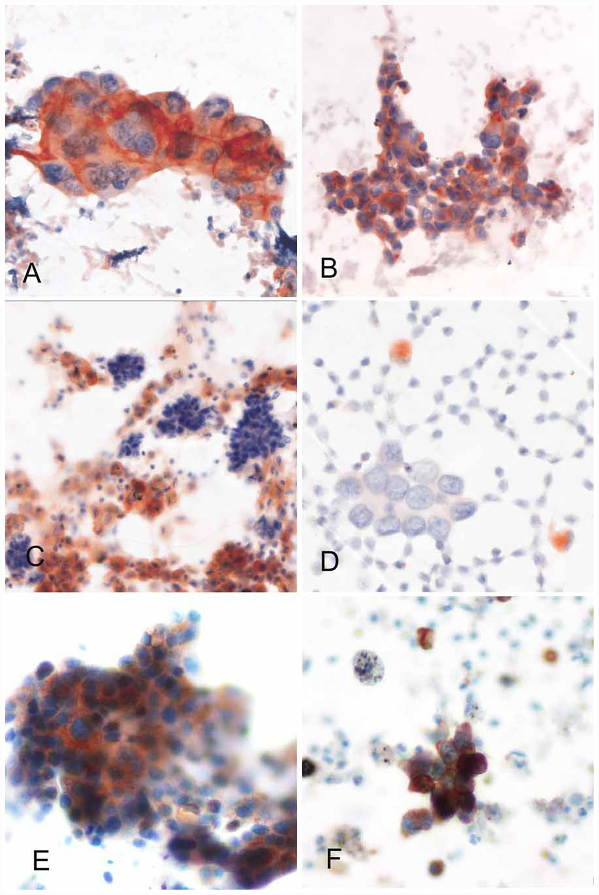
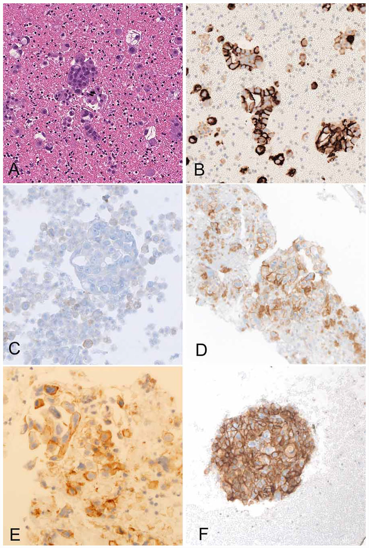
With an escalating number of predictive biomarkers emerging in non-small cell lung carcinoma (NSCLC), immunohistochemistry (IHC) is being used as a rapid and cost-effective tool for the screening and detection of many of these markers. In particular, robust IHC assays performed on formalin-fixed, paraffin-embedded (FFPE) tumor tissue are widely used as surrogate markers for ALK and ROS1 rearrangements and for detecting programmed death ligand 1 (PD-L1) expression in patients with advanced NSCLC; in addition, they have become essential for treatment decisions. Cytology samples represent the only source of tumor in a significant proportion of patients with inoperable NSCLC, and there is increasing demand for predictive biomarker testing on them. However, the wide variation in the types of cytology samples and their preparatory methods, the use of alcohol-based fixatives that interfere with immunochemistry results, the difficulty in procurement of cytology-specific controls, and the uncertainty regarding test validity have resulted in underutilization of cytology material for predictive immunocytochemistry (ICC), and most cytopathologists limit such testing to FFPE cell blocks (CBs). The purpose of this review is to: 1) analyze various preanalytical, analytical, and postanalytical factors influencing ICC results; 2) discuss measures for validation of ICC protocols; and 3) summarize published data on predictive ICC for ALK, ROS1, EGFR gene alterations and PD-L1 expression on lung cancer cytology. Based on our experience and from a review of the literature, we conclude that cytology specimens are in principal suitable for predictive ICC, but proper optimization and rigorous quality control for high-quality staining are essential, particularly for non-CB preparations.
Keywords: cell blocks; immunocytochemistry; lung cancer; predictive; smears.
© 2019 American Cancer Society.
Conflict of interest statement
CONFLICT OF INTEREST DISCLOSURES
Noriko Motoi reports personal fees from Ono pharmaceutical, personal fees from Chugai Pharmaceutical, grants and personal fees from Roche Diagnostics, personal fees from Taiho Pharmaceutical, personal fees from Bristol-Myers Squibb, personal fees from AstraZeneca, personal fees from MSD, personal fees from Novartis, personal fees from Agilent, personal fees from Cook Japan, and personal fees from Miraca Life Sciences. Mauro Papotti reports personal fees from AstraZeneca, personal fees from Roche, personal fees from Pfizer, personal fees from MSD, personal fees from AbbVie, and personal fees from Novartis. Lukas Bubendorf reports grants and personal fees from Roche, grants and personal fees from MSD, personal fees from BMS, personal fees from Astra Zeneca, and personal fees from Pfizer. The other authors have no disclosures.
Figures
Comment in
-
Non-small cell lung cancer predictive biomarker testing via immunocytochemistry: Ways of future past?Cancer Cytopathol. 2019 May;127(5):278-280. doi: 10.1002/cncy.22138. Epub 2019 May 3. Cancer Cytopathol. 2019. PMID: 31050221 No abstract available.
References
-
- Hanna N, Johnson D, Temin S, et al. Systemic therapy for stage IV non-small-cell lung cancer: American Society of Clinical Oncology clinical practice guideline update. J Clin Oncol. 2017;35: 3484–3515. - PubMed
-
- Lindeman NI, Cagle PT, Aisner DL, et al. Updated molecular testing guideline for the selection of lung cancer patients for treatment with targeted tyrosine kinase inhibitors: guideline from the College of American Pathologists, the International Association for the Study of Lung Cancer, and the Association for Molecular Pathology. J Thorac Oncol. 2018;13:323–358. - PubMed
-
- Tsao MS, Kerr KM, Dacic S, Yatabe Y, Hirsch FR, eds. IASLC Atlas of PD-L1 Immunohistochemistry Testing in Lung Cancer. 1st ed Aurora, CO: International Association for the Study of Lung Cancer; 2017.
-
- Thunnissen E, Allen TC, Adam J, et al. Immunohistochemistry of pulmonary biomarkers: a perspective from members of the Pulmonary Pathology Society. Arch Pathol Lab Med. 2018;142: 408–419. - PubMed
Publication types
MeSH terms
Substances
Grants and funding
LinkOut - more resources
Full Text Sources
Medical
Research Materials
Miscellaneous
