. 2019 Jul;13(7):1253-1264.
doi: 10.1002/term.2875. Epub 2019 May 31.

Cardioprotection of PLGA/gelatine cardiac patches functionalised with adenosine in a large animal model of ischaemia and reperfusion injury: A feasibility study

Affiliations

Cardioprotection of PLGA/gelatine cardiac patches functionalised with adenosine in a large animal model of ischaemia and reperfusion injury: A feasibility study

Caterina Cristallini et al. J Tissue Eng Regen Med. 2019 Jul.

Abstract

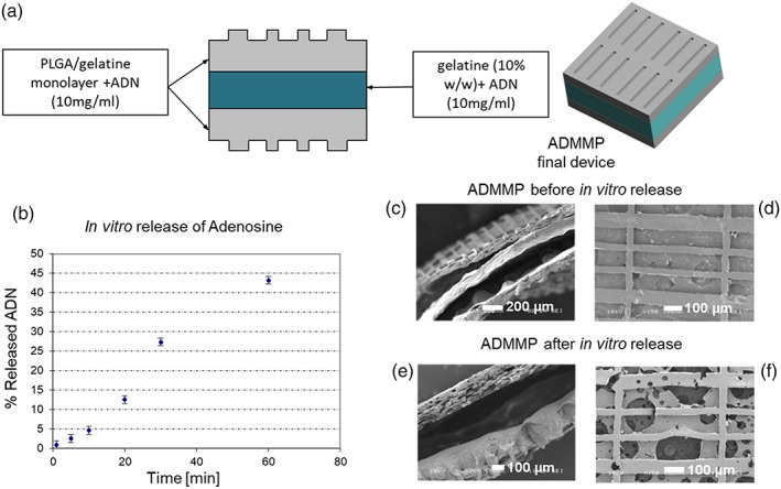
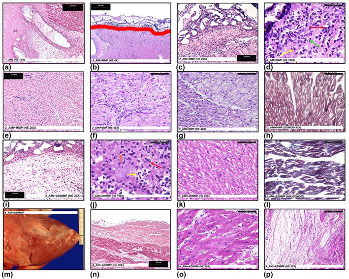
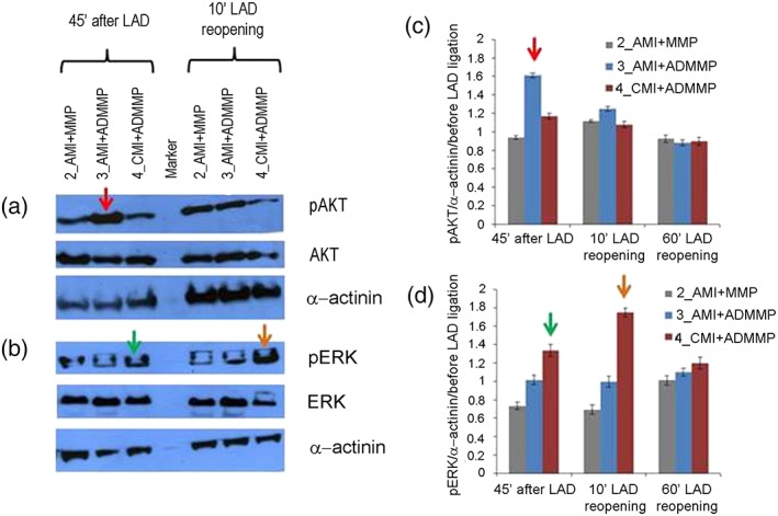
The protection from ischaemia-reperfusion-associated myocardial infarction worsening remains a big challenge. We produced a bioartificial 3D cardiac patch with cardioinductive properties on stem cells. Its multilayer structure was functionalised with clinically relevant doses of adenosine. We report here the first study on the potential of these cardiac patches in the controlled delivery of adenosine into the in vivo ischaemic-reperfused pig heart. A Fourier transform infrared chemical imaging approach allowed us to perform a characterisation, complementary to the histological and biochemical analyses on myocardial samples after in vivo patch implantation, increasing the number of investigations and results on the restricted number of pigs (n = 4) employed in this feasibility step. In vitro tests suggested that adenosine was completely released by a functionalised patch, a data that was confirmed in vivo after 24 hr from patch implantation. Moreover, the adenosine-loaded patch enabled a targeted delivery of the drug to the ischaemic-reperfused area of the heart, as highlighted by the activation of the pro-survival signalling reperfusion injury salvage kinases pathway. At 3 months, though limited to one animal, the used methods provided a picture of a tissue in dynamic conditions, associated to the biosynthesis of new collagen and to a non-fibrotic outcome of the healing process underway. The synergistic effect between the functionalised 3D cardiac patch and adenosine cardioprotection might represent a promising innovation in the treatment of reperfusion injury. As this is a feasibility study, the clinical implications of our findings will require further in vivo investigation on larger numbers of ischaemic-reperfused pig hearts.

Keywords: FT-IR spectroscopy; RISK pathway; adenosine; cardiac patch; cardioprotection; large-animal model.


Conflict of interest statement

The authors declare no conflicts of interest.

Figures

Figure 1
Figure 1
(a) Scheme of ADMMP preparation; (b) release trend of adenosine from the sterilised final scaffold with adenosine loading (ADMMP). SEM images of section and surface of ADMMP (c, d) before and (e, f) after release at 24 hr. The section image of ADMMP before release test shows the thickness of the two microstructured external layers and the thickness of the internal layer containing gelatin and adenosine. The section image after 24‐hr release in MilliQ water points out an intermediate empty space. The surface image of the scaffold shows the presence of free voids, due to the dissolution of the gelatin and adenosine globular structures [Colour figure can be viewed at wileyonlinelibrary.com]
Figure 2
Figure 2
(a) A necrotic myocardium area in 1_AMI (HE 10×); (b) patch adhesion in 2_AMI + MMP. Red line shows the border between patch (up) and epicardium (down) (HE 4×); (c) abundant inflammatory cells under the epicardial layer in 2_AMI + MMP (HE 20×); (d) abundant inflammatory cells under the epicardial layer in 2_AMI + MMP. High magnification (HE 100×). The image shows the presence of macrophages (red arrow) neutrophils (yellow arrow) and eosinophils (green arrow); (e) inflammatory cells inside the myocardium in 2_AMI + MMP (HE 20×); (f) inflammatory cells inside the myocardium in 2_AMI + MMP. High magnification (HE 40×); (g) normal (on the left) and degenerated (on the right) myocardium tissue in 2_AMI + MMP (HE 40×); (h) degenerated myocardium in 2_AMI + MMP (Gomori 40×); (i) inflammatory cells under the epicardial layer in 3_AMI + ADMMP (HE 20×); (j) inflammatory cells under the epicardial layer in 3_AMI + ADMMP. High magnification (HE 100×). The image shows the presence of macrophages (red arrow), neutrophils (yellow arrow), and lymphocytes (orange arrow); (k) suffering myocardium in 3_AMI + ADMMP (HE 40×); (l) suffering myocardium in 3_AMI + ADMMP (Gomori 40×); (m) macroscopic image of the reabsorbed patch in 4_AMI + ADMMP; (n) microscopic image of the reabsorbed patch in 4_AMI + ADMMP (HE 20×); (o) normal myocardium in 4_AMI + ADMMP (HE 40×); (p) mixoid tissue in 4_AMI + ADMMP (HE 20×) [Colour figure can be viewed at wileyonlinelibrary.com]
Figure 3
Figure 3
Analysis of reperfusion injury salvage kinase (RISK) pathway activation: (a, b) western blot analysis of phosphorylated extracellular signal‐regulated kinase AKT (pAKT) and phosphorylated ERK 1/2 (pERK 1/2) normalised to total AKT and ERK 1/2 proteins and to α‐actinin; one representative experiment (out of three) per animal is shown; (c, d) quantification of western blots for pAKT and pERK normalised to α‐actinin and to phosphorylated proteins, detected in the biopsies taken before LAD ligature. SEM of three independent blot quantifications per animal is shown. Differences exceeding ±3 SEM were considered significant and indicated by coloured arrows. A red arrow indicates AKT phosphorylation increase after LAD ligature in animal 3_AMI + ADMMP versus 2_AMI + MMP (1.61 vs. 0.94), a green arrow indicates ERK 1/2 phosphorylation increase after LAD ligature in animal 4_AMI + ADMMP versus 2_AMI + MMP (1.33 versus 0.73), and an orange arrow indicates ERK 1/2 phosphorylation increase 10 min after LAD reopening in animal 4_AMI + ADMMP versus 2_AMI + MMP (1.75 versus 0.69) [Colour figure can be viewed at wileyonlinelibrary.com]
Figure 4
Figure 4
FT‐IR chemical imaging: (a) GAG/protein ratio at the interface with ADMMP in myocardial samples of 3_AMI + ADMMP, (b) GAG/protein ratio in myocardial samples of 2_AMI + MMP. GAG/protein ratio for (c) control healthy tissue, (d) myocardial tissue of 4_AMI + ADMMP in the correspondence of the proximal area, and (e) distal area respect to ADMMP position [Colour figure can be viewed at wileyonlinelibrary.com]
Figure 5
Figure 5
FT‐IR chemical imaging spectra, second derivative spectra, and corresponding chemical map: (a) CH2/CH3 stretching peak ratio for control healthy tissue, (b) for myocardial samples of 4_AMI + ADMMP in the correspondence of proximal area, and (c) distal area respect to ADMMP position [Colour figure can be viewed at wileyonlinelibrary.com]
Figure 6
Figure 6
Second derivative spectra acquired from (a) the control of healthy tissue sections, (b) infarcted area not treated with ADMMP, and (c) proximal area at the interface with ADMMP in 4_AMI + ADMMP [Colour figure can be viewed at wileyonlinelibrary.com]

References

    1. Accomasso, L. , Cristallini, C. , & Giachino, C. (2018). Risk assessment and risk minimization in nanomedicine; a need for predictive, alternative, and 3Rs strategies. Frontiers in Pharmacology, 9, 228 10.3389/fphar.2018.00228 - DOI - PMC - PubMed
    1. Colley, C. S. , Kazarian, S. G. , Weinberg, P. D. , & Lever, M. J. (2004). Spectroscopic imaging of arteries and atherosclerotic plaques. Biopolymers, 74, 328–335. 10.1002/bip.20069 - DOI - PubMed
    1. Cristallini, C. , Cibrario Rocchietti, E. , Accomasso, L. , Folino, A. , Gallina, C. , Muratori, L. , … Giachino, C. (2014). The effect of bioartificial constructs that mimic myocardial structure and biomechanical properties on stem cell commitment towards cardiac lineage. Biomaterials, 35, 92–104. 10.1016/j.biomaterials.2013.09.058 - DOI - PubMed
    1. Cristallini, C. , Cibrario Rocchietti, E. , Gagliardi, M. , Mortati, L. , Saviozzi, S. , Bellotti, E. , … Giachino, C. (2016). Micro and macro‐structured PLGA/gelatin scaffolds promote early cardiogenic commitment of human mesenchymal stem cells in vitro. Stem Cells International, 2016:7176154, 1–16. 10.1155/2016/7176154 - DOI - PMC - PubMed
    1. Donato, M. , D'Annunzio, V. , Berg, G. , Gonzalez, G. , Schreier, L. , Morales, C. , … Gelpi, R. J. (2007). Ischemic postconditioning reduces infarct size by activation of A1 receptors and K+(ATP) channels in both normal and hypercholesterolemic rabbits. Journal of Cardiovascular Pharmacology, 49, 287–292. 10.1097/FJC.0b013e31803c55fe - DOI - PubMed

Publication types

MeSH terms