The Transferrin Receptor CD71 Delineates Functionally Distinct Airway Macrophage Subsets during Idiopathic Pulmonary Fibrosis
- PMID: 31051082
- PMCID: PMC6635794
- DOI: 10.1164/rccm.201809-1775OC
The Transferrin Receptor CD71 Delineates Functionally Distinct Airway Macrophage Subsets during Idiopathic Pulmonary Fibrosis
Abstract
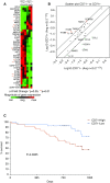
Rationale: Idiopathic pulmonary fibrosis (IPF) is a devastating progressive disease with limited therapeutic options. Airway macrophages (AMs) are key components of the defense of the airways and are implicated in the pathogenesis of IPF. Alterations in iron metabolism have been described during fibrotic lung disease and in murine models of lung fibrosis. However, the role of transferrin receptor 1 (CD71)-expressing AMs in IPF is not known. Objectives: To assess the role of CD71-expressing AMs in the IPF lung. Methods: We used multiparametric flow cytometry, gene expression analysis, and phagocytosis/transferrin uptake assays to delineate the role of AMs expressing or lacking CD71 in the BAL of patients with IPF and of healthy control subjects. Measurements and Main Results: There was a distinct increase in proportions of AMs lacking CD71 in patients with IPF compared with healthy control subjects. Concentrations of BAL transferrin were enhanced in IPF-BAL, and furthermore, CD71- AMs had an impaired ability to sequester transferrin. CD71+ and CD71- AMs were phenotypically, functionally, and transcriptionally distinct, with CD71- AMs characterized by reduced expression of markers of macrophage maturity, impaired phagocytosis, and enhanced expression of profibrotic genes. Importantly, proportions of AMs lacking CD71 were independently associated with worse survival, underlining the importance of this population in IPF and as a potential therapeutic target. Conclusions: Taken together, these data highlight how CD71 delineates AM subsets that play distinct roles in IPF and furthermore show that CD71- AMs may be an important pathogenic component of fibrotic lung disease.
Keywords: airway macrophages; idiopathic pulmonary fibrosis; transferrin receptor.
Figures


Comment in
-
Ironing Out the Roles of Macrophages in Idiopathic Pulmonary Fibrosis.Am J Respir Crit Care Med. 2019 Jul 15;200(2):127-129. doi: 10.1164/rccm.201904-0891ED. Am J Respir Crit Care Med. 2019. PMID: 31091960 Free PMC article. No abstract available.
-
Reply to Puxeddu et al.: CD71- Alveolar Macrophages in Idiopathic Pulmonary Fibrosis: A Look beyond the Borders of the Disease.Am J Respir Crit Care Med. 2019 Dec 1;200(11):1446-1447. doi: 10.1164/rccm.201907-1347LE. Am J Respir Crit Care Med. 2019. PMID: 31347919 Free PMC article. No abstract available.
-
CD71- Alveolar Macrophages in Idiopathic Pulmonary Fibrosis: A Look beyond the Borders of the Disease.Am J Respir Crit Care Med. 2019 Dec 1;200(11):1444-1446. doi: 10.1164/rccm.201906-1159LE. Am J Respir Crit Care Med. 2019. PMID: 31347921 Free PMC article. No abstract available.
References
-
- Martinez FJ, Collard HR, Pardo A, Raghu G, Richeldi L, Selman M, et al. Idiopathic pulmonary fibrosis. Nat Rev Dis Primers. 2017;3:17074. - PubMed
-
- Byrne AJ, Mathie SA, Gregory LG, Lloyd CM. Pulmonary macrophages: key players in the innate defence of the airways. Thorax. 2015;70:1189–1196. - PubMed
-
- Byrne AJ, Maher TM, Lloyd CM. Pulmonary macrophages: a new therapeutic pathway in fibrosing lung disease? Trends Mol Med. 2016;22:303–316. - PubMed
-
- Dancer RCA, Wood AM, Thickett DR. Metalloproteinases in idiopathic pulmonary fibrosis. Eur Respir J. 2011;38:1461–1467. - PubMed

