. 2019 Jul;189(7):1473-1480.
doi: 10.1016/j.ajpath.2019.04.005. Epub 2019 Apr 30.

Choriocapillaris Degeneration in Geographic Atrophy

Affiliations

Choriocapillaris Degeneration in Geographic Atrophy

Elliott H Sohn et al. Am J Pathol. 2019 Jul.

Abstract

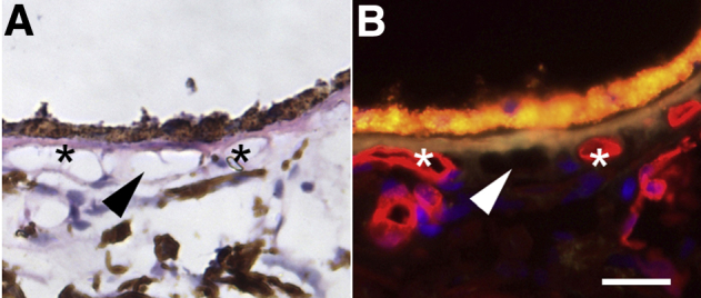
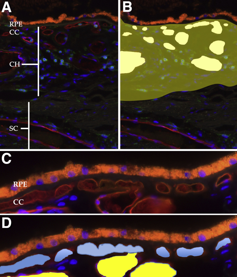
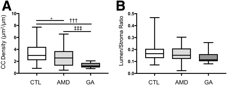
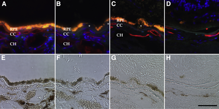
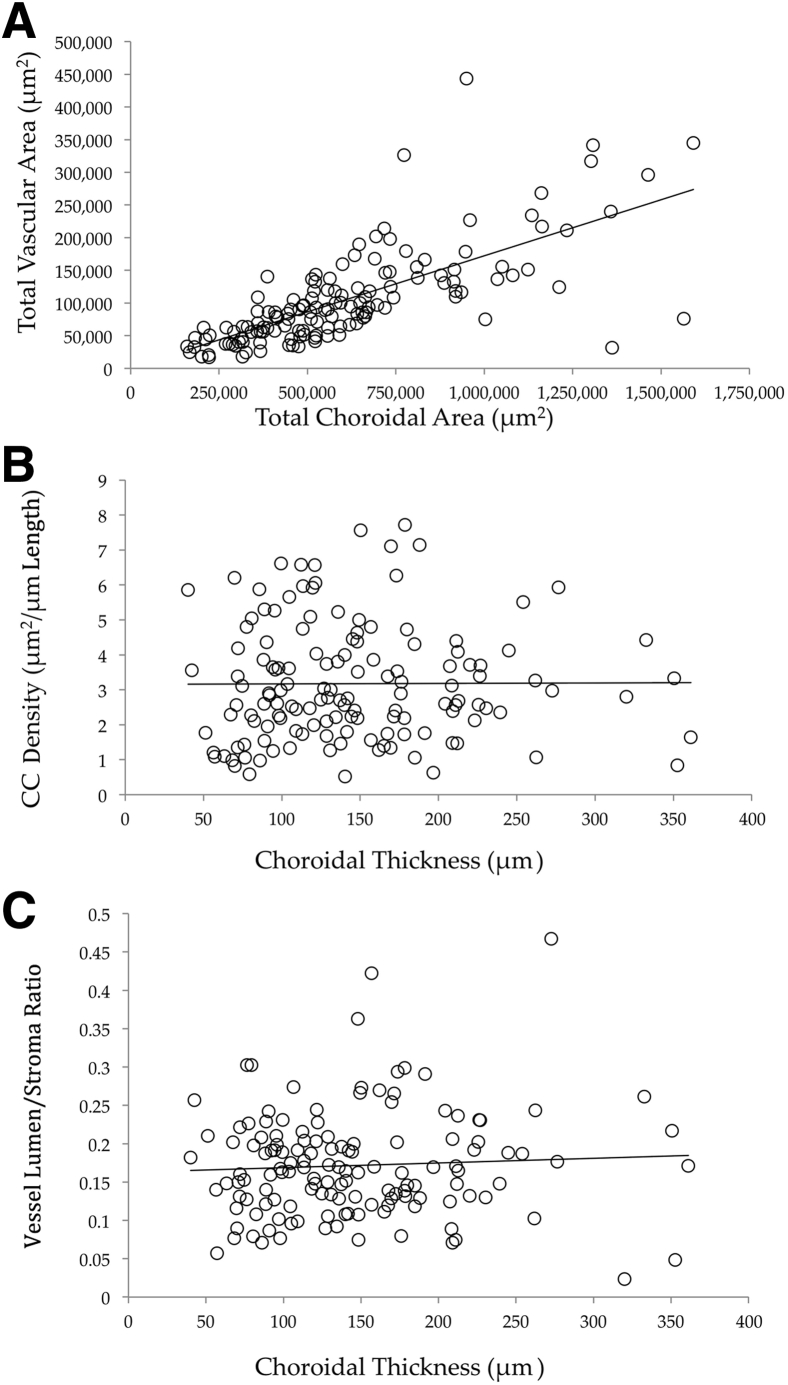
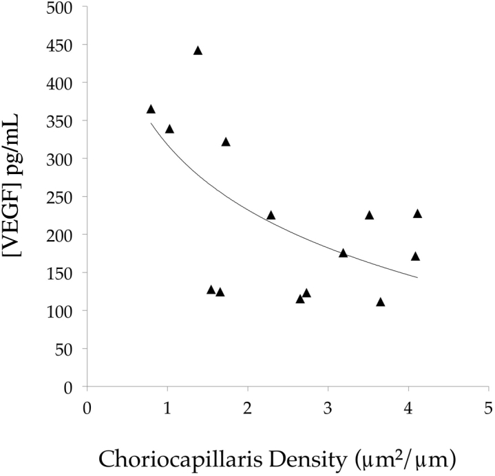
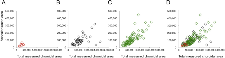
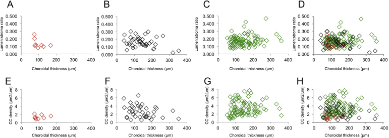
Early age-related macular degeneration (AMD) is characterized by degeneration of the choriocapillaris, the vascular supply of retinal photoreceptor cells. We assessed vascular loss during disease progression in the choriocapillaris and larger vessels in the deeper choroid. Human donor maculae from controls (n = 99), early AMD (n = 35), or clinically diagnosed with geographic atrophy (GA; n = 9, collected from outside the zone of retinal pigment epithelium degeneration) were evaluated using Ulex europaeus agglutinin-I labeling to discriminate between vessels with intact endothelial cells and ghost vessels. Morphometric analyses of choriocapillaris density (cross-sectional area of capillary lumens divided by length) and of vascular lumen/stroma ratio in the outer choroid were performed. Choriocapillaris loss was observed in early AMD (Bonferroni-corrected P = 0.024) with greater loss in GA (Bonferroni-corrected P < 10-9), even in areas of intact retinal pigment epithelium. In contrast, changes in lumen/stroma ratio in the outer choroid were not found to differ between controls and AMD or GA eyes (P > 0.05), suggesting choriocapillaris changes are more prevalent in AMD than those in the outer choroid. In addition, vascular endothelial growth factor-A levels were negatively correlated with choriocapillaris vascular density. These findings support the concept that choroidal vascular degeneration, predominantly in the microvasculature, contributes to dry AMD progression. Addressing capillary loss in AMD remains an important translational target.


Figures

Figure 1
Figure 1
Hematoxylin-eosin–stained section (A) that was previously labeled with Ulex europaeus agglutinin-I (red; B). Section is from a 93-year–old donor with early age-related macular degeneration. The presence of lumina with viable choriocapillaris endothelial cells (asterisks) and ghost vessels (arrowhead) are not readily distinguishable by hematoxylin-eosin staining. Scale bar = 25 μm.
Figure 2
Figure 2
Examples of histologic sections used for quantification of choroidal features. A: Section labeled with Ulex europaeus agglutinin-I lectin (red) for vessel walls and antibodies directed against CD45 (green) for leukocytes, counterstained with DAPI (blue nuclei). The orange autofluorescence is caused by retinal pigment epithelium (RPE) lipofuscin. B: Cross-sectional areas of all vascular elements in the choroid (CH) are quantified (white areas), and vascular areas are divided by total choroidal area (yellow shading) to determine the vascular lumen/stroma ratio. C: Higher-magnification image of the choriocapillaris (CC) and deeper choroidal vessels. D: The segmentation of choriocapillaris (blue) versus larger vessels (yellow). Choriocapillaris lumen areas are measured and divided by length of Bruch membrane to derive the choriocapillaris density. SC, sclera.
Figure 3
Figure 3
Morphometry of 143 donor maculae. A: Choriocapillaris (CC) vascular density (μm2/μm length) is shown for control (CTL), early age-related macular degeneration (AMD), and geographic atrophy (GA) eyes. Choriocapillaris dropout is observed both in early AMD and GA eyes compared with control eyes. B: Choroidal vascular lumen area: stroma area across disease categories. P < 0.05 versus age-related macular degeneration; †††P < 0.001 versus geographic atrophy; ‡‡‡P < 0.001 versus geographic atrophy.
Figure 4
Figure 4
Examples of unaffected control (A and E), early age-related macular degeneration (AMD; B and F), and geographic atrophy (GA; C, D, G, and H) maculae labeled with Ulex europaeus agglutinin-I (UEA-I) lectin (red) and anti-CD45 (green; A–D). Corresponding bright-field images are depicted in E through H. Note the loss of UEA-I–positive capillaries over the course of AMD progression. C and D are from the same macula as G and H, whereas C and G are outside of the central atrophic lesion depicted in D and H. Donor ages are 86, 82, and 99 years, respectively. Asterisks indicate drusen. Scale bar = 50 μm. CC, choriocapillaris; CH, outer choroid; RPE, retinal pigment epithelium.
Figure 5
Figure 5
Vascular parameter relationship with choroidal thickness. A: Total vascular lumen area, plotted against total choroidal area, shows an expected relationship between these parameters. B and C: Choriocapillaris (CC) density (μm2/μm length; B) and outer choroid (CH) lumen/stroma ratio (C) were plotted against choroidal thickness (μm). Neither CC nor CH vessel density is associated with choroidal thickness. Solid lines show regression lines.
Figure 6
Figure 6
Relationship between choriocapillaris vascular density and retinal vascular endothelial growth factor (VEGF) concentration. In eyes with a low vascular density, VEGF levels are increased, consistent with retinal hypoxia overlying lost choroidal vasculature. Solid line shows trend line.
Supplemental Figure S1
Plots depicting total choroidal area (x axis) versus outer choroid vascular lumen areas (y axis) in different disease states. All values are given in μm2. A: Eyes with geographic atrophy (GA). B: Eyes with early age-related macular degeneration (AMD). C: Control eyes. D: Overlay of A through C. Slopes for GA, early AMD, and controls are 0.117, 0.104, and 0.202, respectively. Total measured choroidal area is lowest in GA eyes because of the thinner choroids in these samples.
Supplemental Figure S2
Relationship between choroidal thickness (x axis) and large-vessel lumen/stroma ratios (A–D) and choriocapillaris (CC) vascular density (E–H), shown for geographic atrophy eyes (A and E), early age-related macular degeneration eyes (B and F), unaffected, age-matched controls (C and G), and merged panels (D and H).

References

    1. Nickla D.L., Wallman J. The multifunctional choroid. Prog Retin Eye Res. 2010;29:144–168. - PMC - PubMed
    2. Nickla DL, Wallman J: The multifunctional choroid. Prog Retin Eye Res 2010, 29:144-168 - PMC - PubMed
    1. Chirco K.R., Sohn E.H., Stone E.M., Tucker B.A., Mullins R.F. Structural and molecular changes in the aging choroid: implications for age-related macular degeneration. Eye (Lond) 2017;31:10–25. - PMC - PubMed
    2. Chirco KR, Sohn EH, Stone EM, Tucker BA, Mullins RF: Structural and molecular changes in the aging choroid: implications for age-related macular degeneration. Eye (Lond) 2017, 31:10-25 - PMC - PubMed
    1. Linsenmeier R.A., Braun R.D. Oxygen distribution and consumption in the cat retina during normoxia and hypoxemia. J Gen Physiol. 1992;99:177–197. - PMC - PubMed
    2. Linsenmeier RA, Braun RD: Oxygen distribution and consumption in the cat retina during normoxia and hypoxemia. J Gen Physiol 1992, 99:177-197 - PMC - PubMed
    1. Harris A. Butterworth Heinemann; Philadelphia, PA: 2003. Atlas of Ocular Blood Flow.
    2. Harris A: Atlas of Ocular Blood Flow. 2003,
    1. Whitmore S.S., Sohn E.H., Chirco K.R., Drack A.V., Stone E.M., Tucker B.A., Mullins R.F. Complement activation and choriocapillaris loss in early AMD: implications for pathophysiology and therapy. Prog Retin Eye Res. 2014;45:1–29. - PMC - PubMed
    2. Whitmore SS, Sohn EH, Chirco KR, Drack AV, Stone EM, Tucker BA, Mullins RF: Complement activation and choriocapillaris loss in early AMD: Implications for pathophysiology and therapy. Prog Retin Eye Res 2014, 45:1-29 - PMC - PubMed

Publication types

Substances