. 2019 Apr 30;9(2):48.
doi: 10.3390/diagnostics9020048.

HAMP Downregulation Contributes to Aggressive Hepatocellular Carcinoma via Mechanism Mediated by Cyclin4-Dependent Kinase-1/STAT3 Pathway

Affiliations

HAMP Downregulation Contributes to Aggressive Hepatocellular Carcinoma via Mechanism Mediated by Cyclin4-Dependent Kinase-1/STAT3 Pathway

Ying Shen et al. Diagnostics (Basel). .

Abstract

Background: Hepcidin encoded by HAMP is vital to regulating proliferation, metastasis, and migration. Hepcidin is secreted specifically by the liver. This study sought to examine the functional role of hepcidin in hepatocellular carcinoma (HCC).

Methods: Data in the Cancer Genome Atlas database was used to analyze HAMP expression as it relates to HCC prognosis. We then used the 5-ethynyl-20-deoxyuridine (EdU) incorporation assay, transwell assay, and flow cytometric analysis, respectively, to assess proliferation, migration, and the cell cycle. Gene set enrichment analysis (GSEA) was used to find pathways affected by HAMP.

Results: HAMP expression was lower in hepatocellular carcinoma samples compared with adjacent normal tissue controls. Low HAMP expression was linked with a higher rate of metastasis and poor disease-free status. Downregulation of HAMP induced SMMC-7721 and HepG-2 cell proliferation and promoted their migration. HAMP could affect the cell cycle pathway and Western blotting, confirming that reduced HAMP levels activated cyclin-dependent kinase-1/stat 3 pathway.

Conclusion: Our findings indicate that HAMP functions as a tumor suppressor gene. The role of HAMP in cellular proliferation and metastasis is related to cell cycle checkpoints. HAMP could be considered as a diagnostic biomarker and targeted therapy in HCC.

Keywords: HAMP; cell cycle; hepatocellular carcinoma; iron; metastasis.


Conflict of interest statement

The authors declare no conflict of interests.

Figures

Figure 1
Figure 1
HAMP expression is reduced in liver hepatocellular carcinoma (HCC) and low HAMP expression is linked to higher cancer metastasis stage code and poor disease-free status. (A) HAMP expression in HCC tumors was significantly decreased relative to adjacent liver tissue (p < 0.0001). (B) Histogram showing HAMP expression in liver hepatocellular cancer. The 2−ΔΔCt approach was used to calculate HAMP expression, and expression in each patient is given as the tumor (T, n = 50)/normal (N, n = 50) ratio; (C) Low HAMP expression is associated with high cancer metastasis in HCC patients (p = 0.0121). (D) Low HAMP expression is associated with poor disease-free status in HCC patients, according to a Kaplan–Meier analysis of HCC (n = 320, log rank test, p = 0.0431).
Figure 2
Figure 2
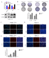
The effect of HAMP on proliferation. (A) Analysis of HAMP expression in SMMC-7721/HepG-2 cells after transfection with control, HAMP shRNA, and HAMP overexpression vector. (B) A tumor colony forming assay was used to detect the proliferation of SMMC-7721 and HepG-2 cells after knockdown or overexpressed the HAMP gene expression. (CF) 5-ethynyl-20-deoxyuridine (EdU) incorporation assay was employed to detect the viability of SMMC-7721/HepG-2 cells transfected with control, HAMP shRNA, and HAMP overexpression vector. # p < 0.05. Scale bar = 50 µM
Figure 3
Figure 3
The effect of HAMP on the migration of liver cancer cells. (A,B) Migration was measured via wound-healing assay for SMMC-7721/HepG-2 cells transfected with control, HAMP shRNA, and HAMP overexpression vector. # p < 0.05. Scale bar = 300 µM
Figure 4
Figure 4
HAMP affects hepatocellular carcinoma proliferation in vivo. SMMC-7721 cells transfected with control or shRNA were injected s.c. in BALB/c-nu mice. (A) HAMP expression in tumors. (B) Tumor volume, measured every other day. (C,D) Tumor volumes changes in control and shRNA mice. (E) Tumor weight changes in control and shRNA mice. (n = 5). # p < 0.05.
Figure 5
Figure 5
Gene set enrichment analysis (GSEA) based on HAMP expression in HCC patients. To identify pathways linked to HAMP expression in The Cancer Genome Atlas (TCGA) samples, we compared gene expression in those with low HAMP expression (blue) and high HAMP expression (red). Visualizations were produced using Cytoscape and Enrichment map (1% FDR, p < 0.005). Each node is representative of a set of enriched genes, with annotations being ascribed based on how similar the sets are to one another. A network of nodes was used to map the enrichment results, with the size of nodes being directly correlated with the gene number in that particular gene set. The number of genes shared between two given gene sets determined the thickness of the green line connecting them. The final map was generated via removal of any general or noninformative smaller networks in order to simplify the final diagram. (A) Go enrichment plot showed HAMP downregulation is related to cell cycle checkpoint. (B) KEGG enrichment plot showed HAMP downregulation is related to the cell cycle.
Figure 6
Figure 6
HAMP downregulation activates the cdk1/stat3 pathway. (A) Knockdown of HAMP promotes cellular iron concentration in SMMC-7721/HepG-2 cells. (B) Cell cycle analysis of SMMC-7721 and HepG-2 cells transfected with HAMP shRNA. (C,D) HAMP downregulation activates the cdk1/stat3 pathway. (E) Downregulation of HAMP low expression of hepcidin, which promotes cellular iron concentration and then activates the cdk1/stat3 pathway to promote tumor proliferation and metastasis. # p < 0.05, * p < 0.05 and ** p < 0.01.

Similar articles

Cited by

References

    1. Sia D., Villanueva A., Friedman S.L., Llovet J.M. Liver Cancer Cell of Origin, Molecular Class, and Effects on Patient Prognosis. Gastroenterology. 2017;152:745–761. doi: 10.1053/j.gastro.2016.11.048. - DOI - PubMed
    1. Lin M.V., King L.Y., Chung R.T. Hepatitis C virus-associated cancer. Annu. Rev. Pathol. 2015;10:345–370. doi: 10.1146/annurev-pathol-012414-040323. - DOI - PubMed
    1. Llovet J.M., Villanueva A., Lachenmayer A., Finn R.S. Advances in targeted therapies for hepatocellular carcinoma in the genomic era. Nat. Rev. Clin. Oncol. 2015;12:408–424. doi: 10.1038/nrclinonc.2015.103. - DOI - PubMed
    1. Talwalkar J.A., Gores G.J. Diagnosis and staging of hepatocellular carcinoma. Gastroenterology. 2004;127:S126–S132. doi: 10.1053/j.gastro.2004.09.026. - DOI - PubMed
    1. Kim C.K., He P., Bialkowska A.B., Yang V.W. SP and KLF Transcription Factors in Digestive Physiology and Diseases. Gastroenterology. 2017;152:1845–1875. doi: 10.1053/j.gastro.2017.03.035. - DOI - PMC - PubMed

LinkOut - more resources