. 2019 Apr 30;17(5):258.
doi: 10.3390/md17050258.

Effects of Fucoidans from Five Different Brown Algae on Oxidative Stress and VEGF Interference in Ocular Cells

Affiliations

Effects of Fucoidans from Five Different Brown Algae on Oxidative Stress and VEGF Interference in Ocular Cells

Philipp Dörschmann et al. Mar Drugs. .

Abstract

Background: Fucoidans are interesting for potential usage in ophthalmology, and especially age-related macular degeneration. However, fucoidans from different species may vary in their effects. Here, we compare fucoidans from five algal species in terms of oxidative stress protection and vascular endothelial growth factor (VEGF) interference in ocular cells.

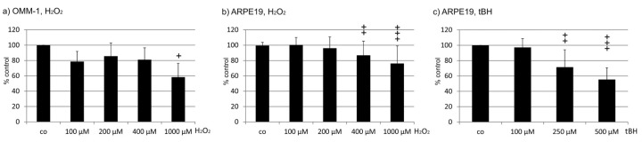
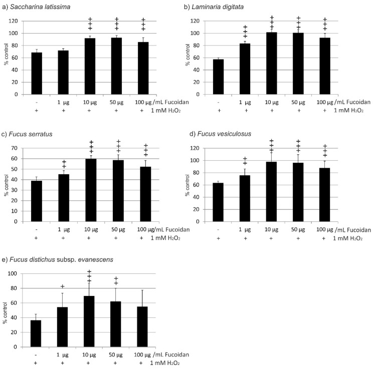
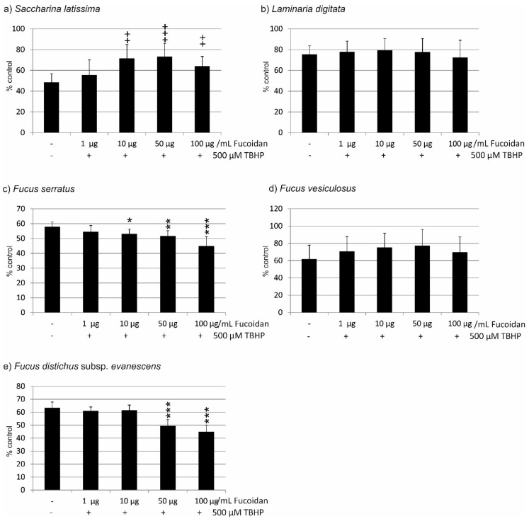
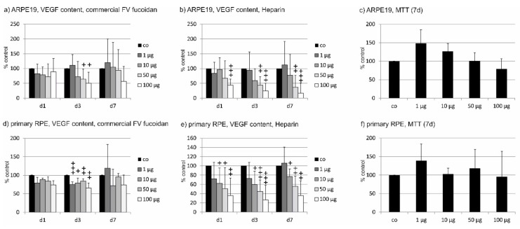
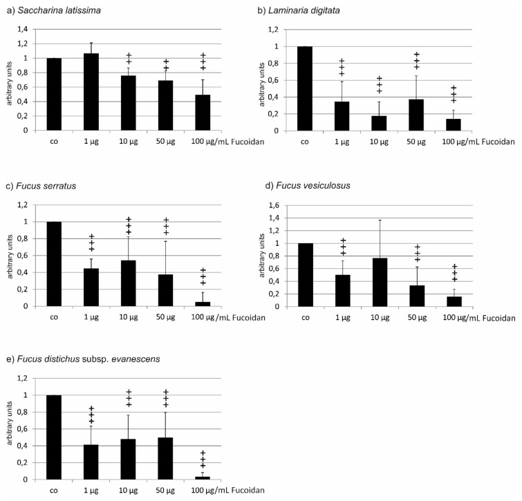
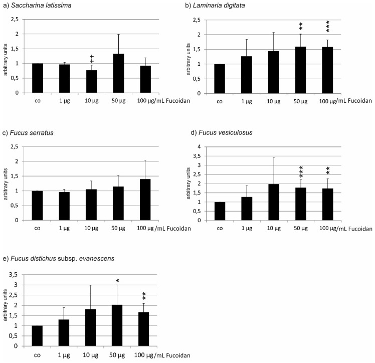
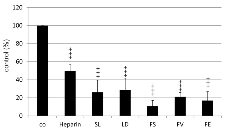
Methods: Brown algae (Fucus vesiculosus, Fucus distichus subsp. evanescens, Fucus serratus, Laminaria digitata, Saccharina latissima) were harvested and fucoidans isolated by hot-water extraction. Fucoidans were tested in several concentrations (1, 10, 50, and 100 µg/mL). Effects were measured on a uveal melanoma cell line (OMM-1) (oxidative stress), retinal pigment epithelium (RPE) cell line ARPE19 (oxidative stress and VEGF), and primary RPE cells (VEGF). Oxidative stress was induced by H2O2 or tert-Butyl hydroperoxide (TBHP). Cell viability was investigated with methyl thiazolyl tetrazolium (MTT or MTS) assay, and VEGF secretion with ELISA. Affinity to VEGF was determined by a competitive binding assay.

Results: All fucoidans protected OMM-1 from oxidative stress. However, in ARPE19, only fucoidan from Saccharina latissima was protective. The affinity to VEGF of all fucoidans was stronger than that of heparin, and all reduced VEGF secretion in ARPE19. In primary RPE, only the fucoidan from Saccharina latissima was effective.

Conclusion: Among the fucoidans from five different species, Saccharina latissima displayed the most promising results concerning oxidative stress protection and reduction of VEGF secretion.

Keywords: Fucus distichus subsp. evanescens; Fucus serratus; Fucus vesiculosus; Laminaria digitata; Saccharina latissima; VEGF; age-related macular degeneration; fucoidan; oxidative stress.


Conflict of interest statement

The authors declare no conflict of interest.

Figures

Figure 1
Figure 1
Characterization of the susceptibility of cell lines to oxidative stress. Cell viability was tested in OMM-1 (a) and ARPE19 (b) exposed to H2O2 (a,b) and tert-Butyl hydroperoxide (TBHP) (c). Significance was evaluated with Friedman’s ANOVA and Student’s t-test, + p < 0.05, ++ p < 0.01, +++ p < 0.001 compared to control (n > 3).
Figure 2
Figure 2
Cell viability of OMM-1 cells challenged with 1 mM H2O2 after incubation with fucoidan from (a) Saccharina latissima (SL), (b) Laminaria digitata (LD), (c) Fucus serratus (FS), (d) Fucus vesiculosus (FV), (e) Fucus distichus subsp. evanescens (FE). Cell viability was measured by MTS assay and is depicted as mean and standard deviation, with the control set as 100%. All fucoidans tested displayed protective effects, with the efficacy of LD > FV > SL > FE > FS. Significance was evaluated with Friedman’s ANOVA and subsequent Student’s t-test, + p < 0.05, ++ p < 0.01, +++ p < 0.001, all versus 1 mM H2O2 (n = 8).
Figure 3
Figure 3
Cell viability of ARPE19 cells challenged with 500 µM TBHP after incubation with fucoidan from (a) Saccharina latissima (SL), (b) Laminaria digitata (LD), (c) Fucus serratus (FS), (d) Fucus vesiculosus (FV), (e) Fucus distichus subsp. evanescens (FE). Cell viability was measured by MTS assay and is depicted as mean and standard deviation, with the control set as 100%. Only SL fucoidan displayed a protective effect, while FS and FE fucoidans reduced cell viability. Significance was evaluated with Friedman’s ANOVA and subsequent Student’s t-test, ++ p < 0.01, +++ p < 0.001, for protective effects against 500 µM TBHP, * p < 0.05, ** p < 0.01 and *** p < 0.001 for exacerbating effects against 500 µM TBHP (n = 8).
Figure 4
Figure 4
Effect of incubation time for vascular endothelial growth factor (VEGF) experiments. Commercially available fucoidan from Fucus vesiculosus was applied for 1 day (d1), 3 days (d3), and 7 days (d7), respectively, at indicated concentrations, to ARPE19 (a) or primary porcine retinal pigment epithelium (RPE) (d). In addition, heparin was tested on ARPE19 (b) and RPE cells (e). Cell viability was tested after 7 days (c,f). VEGF reduction was primarily seen after 3 days of incubation. Heparin showed a dose-dependent effect with similar significant reductions at all tested time points for concentrations of 10–100 µg/mL. No toxic effect was seen after 7 days in either cell type. Significance was evaluated with Friedman’s ANOVA and subsequent Student’s t-test, + p < 0.05 ++ p < 0.01, +++ p < 0.001 against the control (n = 4–6).
Figure 5
Figure 5
VEGF secretion in ARPE19 cells after incubation with different concentrations of fucoidan. (a) Saccharina latissima (SL), (b) Laminaria digitata (LD), c) Fucus serratus (FS), (d) Fucus vesiculosus (FV), (e) Fucus distichus subsp. evanescens (FE). VEGF content was evaluated by ELISA and normalized to cell viability. Control = 1. In ARPE19 cells, all fucoidans reduced VEGF content with the efficacy of LD > FS > FE > FV > SL. Significance was evaluated with Friedman’s ANOVA and subsequent Student’s t-test, ++ p < 0.01, +++ p < 0.001 compared to the control (n = 6–8).
Figure 6
Figure 6
VEGF secretion of primary porcine RPE cells after incubation with different concentrations of fucoidan from (a) Saccharina latissima (SL), (b) Laminaria digitata (LD), (c) Fucus serratus (FS), (d) Fucus vesiculosus (FV), (e) Fucus distichus subsp. evanescens (FE). VEGF content was evaluated in ELISA and normalized to cell viability. Control = 1. In RPE cells, only SL fucoidan reduced the VEGF content of RPE cells (10 µg/mL), while LD, FV, and FE induced a higher signal at concentrations of 50 and 100 µg/mL. Significance was evaluated with Friedman’s ANOVA and subsequent Student’s t-test, + p < 0.05 reduction compared to the control, * p < 0.05, ** p < 0.01 and *** p < 0.001 (n = 7).
Figure 7
Figure 7
VEGF binding affinity of fucoidans from Saccharina latissima (SL), Laminaria digitata (LD), Fucus serratus (FS), Fucus vesiculosus (FV), Fucus distichus subsp. evanescens (FE), and of heparin. Significance was evaluated with Friedman’s ANOVA and subsequent Student’s t-test, +++ p < 0.001 compared to control (biotinylated heparin).

References

    1. Li B., Lu F., Wei X., Zhao R. Fucoidan: Structure and bioactivity. Molecules. 2008;13:1671–1695. doi: 10.3390/molecules13081671. - DOI - PMC - PubMed
    1. Fitton J.H., Stringer D.N., Karpiniec S.S. Therapies from Fucoidan: An Update. Mar. Drugs. 2015;13:5920–5946. doi: 10.3390/md13095920. - DOI - PMC - PubMed
    1. Dithmer M., Fuchs S., Shi Y., Schmidt H., Richert E., Roider J., Klettner A. Fucoidan reduces secretion and expression of vascular endothelial growth factor in the retinal pigment epithelium and reduces angiogenesis in vitro. PLoS ONE. 2014;9:e89150. doi: 10.1371/journal.pone.0089150. - DOI - PMC - PubMed
    1. Klettner A. Fucoidan as a potential therapeutic for major blinding diseases—A hypothesis. Mar. Drugs. 2016;14:E31. doi: 10.3390/md14020031. - DOI - PMC - PubMed
    1. Miller J.W. Age-related macular degeneration revisited--piecing the puzzle: The LXIX Edward Jackson memorial lecture. Am. J. Ophthalmol. 2013;155:1–35. doi: 10.1016/j.ajo.2012.10.018. - DOI - PubMed

LinkOut - more resources