Review
. 2019 Apr 30;8(5):401.
doi: 10.3390/cells8050401.

Mitochondrial Flexibility of Breast Cancers: A Growth Advantage and a Therapeutic Opportunity

Affiliations
Review

Mitochondrial Flexibility of Breast Cancers: A Growth Advantage and a Therapeutic Opportunity

Angelica Avagliano et al. Cells. .

Abstract

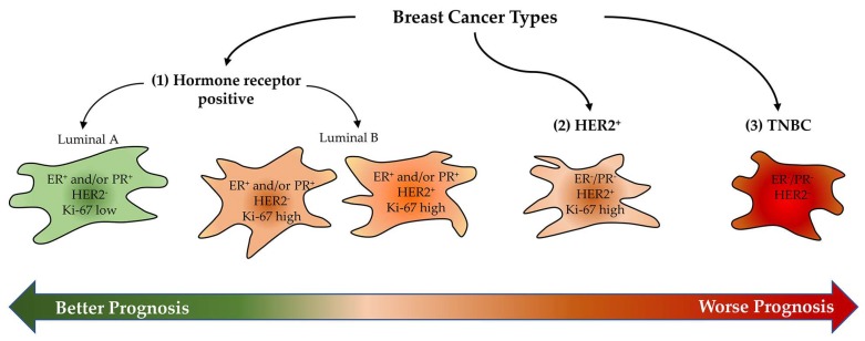
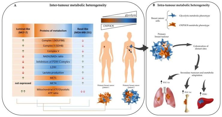
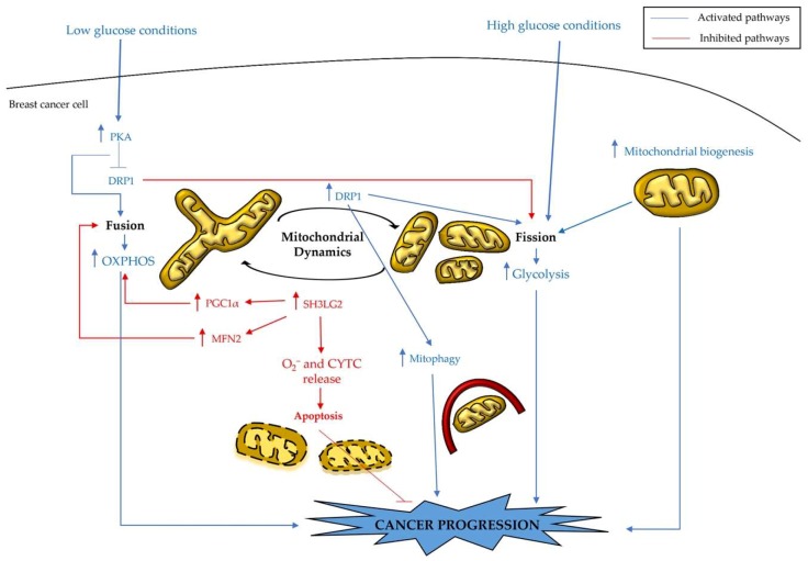
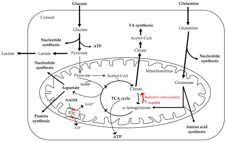
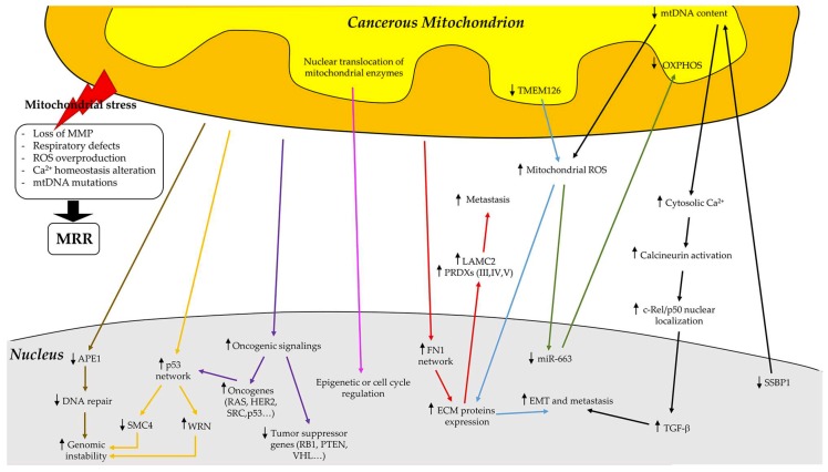
Breast cancers are very heterogeneous tissues with several cell types and metabolic pathways together sustaining the initiation and progression of disease and contributing to evasion from cancer therapies. Furthermore, breast cancer cells have an impressive metabolic plasticity that is regulated by the heterogeneous tumour microenvironment through bidirectional interactions. The structure and accessibility of nutrients within this unstable microenvironment influence the metabolism of cancer cells that shift between glycolysis and mitochondrial oxidative phosphorylation (OXPHOS) to produce adenosine triphosphate (ATP). In this scenario, the mitochondrial energetic pathways of cancer cells can be reprogrammed to modulate breast cancer's progression and aggressiveness. Moreover, mitochondrial alterations can lead to crosstalk between the mitochondria and the nucleus, and subsequently affect cancer tissue properties. This article reviewed the metabolic plasticity of breast cancer cells, focussing mainly on breast cancer mitochondrial metabolic reprogramming and the mitochondrial alterations influencing nuclear pathways. Finally, the therapeutic strategies targeting molecules and pathways regulating cancer mitochondrial alterations are highlighted.

Keywords: breast cancer; mitochondrial reprogramming; oxidative phosphorylation; therapeutic strategies; tumour microenvironment.


Conflict of interest statement

The authors declare no conflict of interest.

Figures

Figure 1
Figure 1
Classification of breast cancer into three major types based on their immunohistochemical properties and relative prognosis.
Figure 2
Figure 2
Breast cancer as a heterogeneous metabolic disease. (A) Inter-tumour metabolic heterogeneity refers to the ability of different breast cancer types to exhibit a distinct and preferential metabolic phenotype. MCF-7 cells, belonging to the luminal-like breast cancer subtype, are more dependent on mitochondrial respiration, and reduce lactate dehydrogenase (LDH) expression, and by consequence lactate production. MCF-7 cells increase monocarboxylate transporter 1 (MCT-1) protein levels to import the lactate produced by the tumour microenvironment into the cell. Conversely, the basal-like MDA-MB-231 cells exhibit a more glycolytic phenotype. The lower respiratory rate in MDA-MB-231 cells is associated with a strong decrease in complexes I, II, and V of the electron transport chain (ETC). As a result of dysfunctional Complex I, MDA-MB-231 cells exhibit higher levels of NADH, which in turn inhibits the pyruvate dehydrogenase (PDH) complex. Thus, pyruvate does not enter the tricarboxylic acid (TCA) cycle, but it is mainly converted to lactate by LDH, which is highly expressed in MDA-MB-231 cells. Lactate is efficiently extruded from MDA-MB-231 cells through the monocarboxylate transporter 4 (MCT-4), which is not expressed in MCF-7 cells. (B) Intra-tumour metabolic heterogeneity refers to the presence in the same tumour mass of a heterogeneous cell population displaying different metabolic phenotypes. Breast cancer cells are able to adapt their metabolism, according to several stresses and signals sent by the ever-changing and harsh microenvironment of both primary tumour and metastatic sites. In liver metastasis, breast cancer cells are more glycolytic and express high levels of pyruvate dehydrogenase kinase 1 (PDK1). In bone and lung metastasis, breast cancer cells preferentially use mitochondrial oxidative phosphorylation (OXPHOS) and upregulate peroxisome proliferator-activated receptor gamma coactivator-1 alpha (PGC-1α).
Figure 3
Figure 3
Mitochondrial morphology alterations and metabolic reprogramming. Mitochondrial morphology transitions (from elongated to fragmented, and vice versa) are linked to breast cancer metabolism. In fact, breast cancer cells alter mitochondrial dynamics to adjust their bioenergetics and biosynthetic needs in order to survive in harsh conditions and support tumour progression. In high-nutrient conditions, mitochondria are fragmented and dysfunctional, and energy demand is mainly supported by glycolysis. Dynamin-related protein 1 (DRP1) upregulation in breast cancer cells is associated with fission, glycolysis, and mitophagy. The reduction of mitochondrial number due to mitophagy is restored by an increase in mitochondrial biogenesis. The fission mechanism is also induced in breast cancer by the loss of the tumour suppressor gene, SH3LG2. When overexpressed, endophilin-A1 encoded by the SH3LG2 gene translocates to mitochondria, interacts with PGC-1α and Mitofusin 2 (MFN2), and triggers the mitochondrial fusion network and OXPHOS. Moreover, the mitochondrial translocation of SH3GL2 triggers the intrinsic apoptotic pathway through the induction of O2 production and cytochrome C (CYTC) release. Conversely, in low nutrient conditions, breast cancer cells maintain their mitochondria in a hyperfused state by inhibiting the mitochondrial fission protein DRP1 and favouring energy production through OXPHOS. The blue arrows indicate activated pathways, whereas red arrows depict inhibited pathways in the breast cancer cell.
Figure 4
Figure 4
Functional alterations of mitochondria in breast cancer cells. Glucose carbon, which is mainly used for lactate production, is directed away from the TCA cycle and fatty acid (FA) synthesis. In this context, glutamine carbon feeds the TCA cycle and contributes to FA synthesis through the reductive pathway. The reductive carboxylation is supported by a disturbance in the redox ratio of mitochondria due to the aberrant function of the ETC, which decreases the NAD+/NADH ratio. This ratio is dissipated in part through the transfer of reducing equivalents from NADH to NADPH, which in turn may drive the NADPH-dependent reductive carboxylation of glutamine by isocitrate dehydrogenase 1 (IDH1) and IDH2. All these processes are further enhanced during hypoxia. Moreover, cells with ETC dysfunction use pyruvate, which is formed during glycolysis, to produce aspartate, which is important for cell proliferation and survival.
Figure 5
Figure 5
Mitochondrial retrograde regulation (MRR). Schematic representation of the main pathways used by cancerous mitochondria to communicate with the nucleus during breast cancer progression.

References

    1. Ruocco M.R., Avagliano A., Granato G., Imparato V., Masone S., Masullo M., Nasso R., Montagnani S., Arcucci A. Involvement of Breast Cancer-Associated Fibroblasts in Tumor Development, Therapy Resistance and Evaluation of Potential Therapeutic Strategies. Curr. Med. Chem. 2018;25:3414–3434. doi: 10.2174/0929867325666180309120746. - DOI - PubMed
    1. Arcucci A., Ruocco M.R., Granato G., Sacco A.M., Montagnani S. Cancer: An Oxidative Crosstalk between Solid Tumor Cells and Cancer Associated Fibroblasts. Biomed. Res. Int. 2016;2016:4502846. doi: 10.1155/2016/4502846. - DOI - PMC - PubMed
    1. Sotgia F., Ozsvari B., Fiorillo M., De Francesco E.M., Bonuccelli G., Lisanti M.P. A mitochondrial based oncology platform for targeting cancer stem cells (CSCs): MITO-ONC-RX. Cell Cycle. 2018;17:2091–2100. doi: 10.1080/15384101.2018.1515551. - DOI - PMC - PubMed
    1. Martinez-Outschoorn U.E., Peiris-Pagés M., Pestell R.G., Sotgia F., Lisanti M.P. Cancer metabolism: A therapeutic perspective. Nat. Rev. Clin. Oncol. 2017;14:11–31. doi: 10.1038/nrclinonc.2016.60. - DOI - PubMed
    1. Avagliano A., Granato G., Ruocco M.R., Romano V., Belviso I., Carfora A., Montagnani S., Arcucci A. Metabolic Reprogramming of Cancer Associated Fibroblasts: The Slavery of Stromal Fibroblasts. Biomed. Res. Int. 2018;2018:6075403. doi: 10.1155/2018/6075403. - DOI - PMC - PubMed

Publication types