. 2019 Apr 30;11(5):602.
doi: 10.3390/cancers11050602.

Hypoxia Selectively Impairs CAR-T Cells In Vitro

Affiliations

Hypoxia Selectively Impairs CAR-T Cells In Vitro

Robert Berahovich et al. Cancers (Basel). .

Abstract

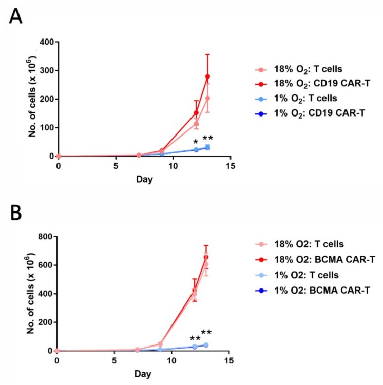
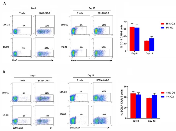
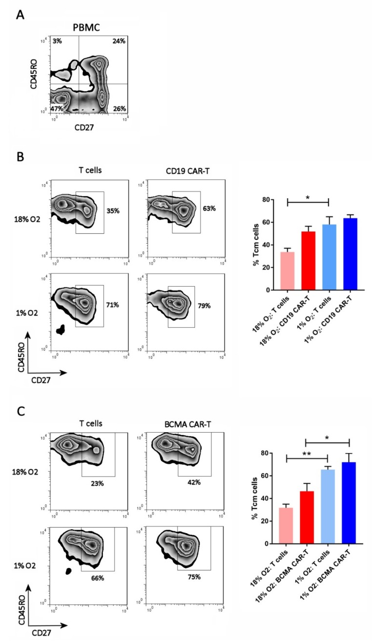
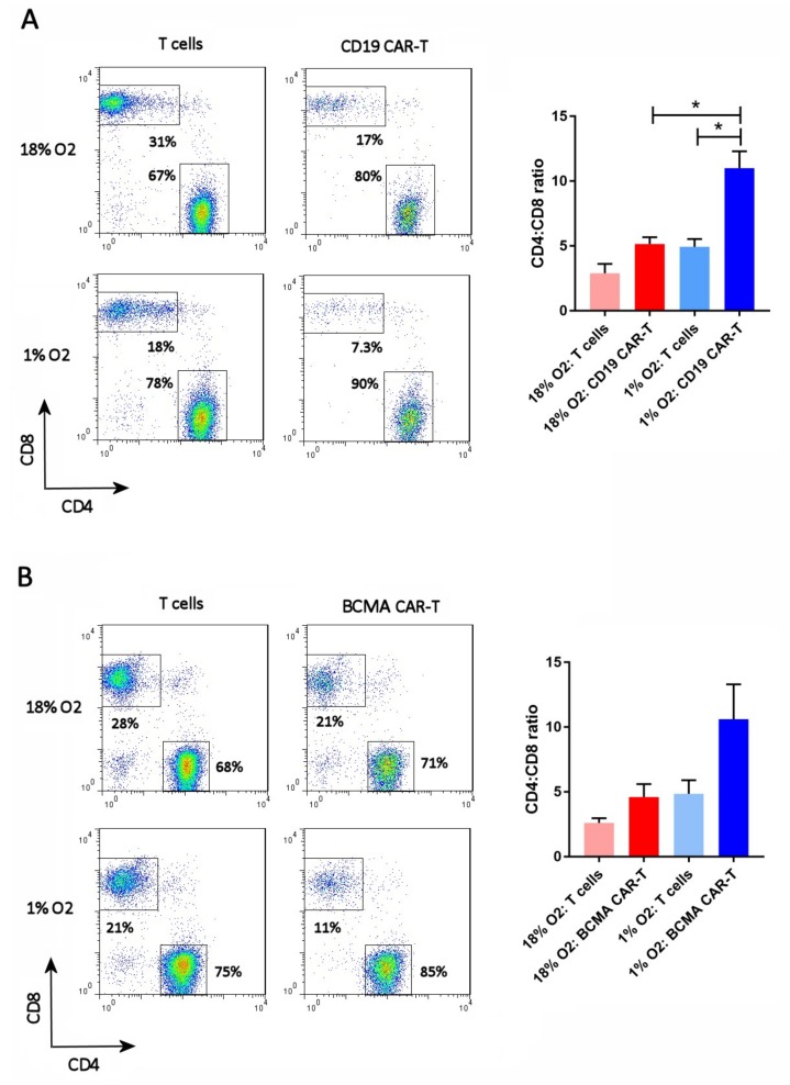
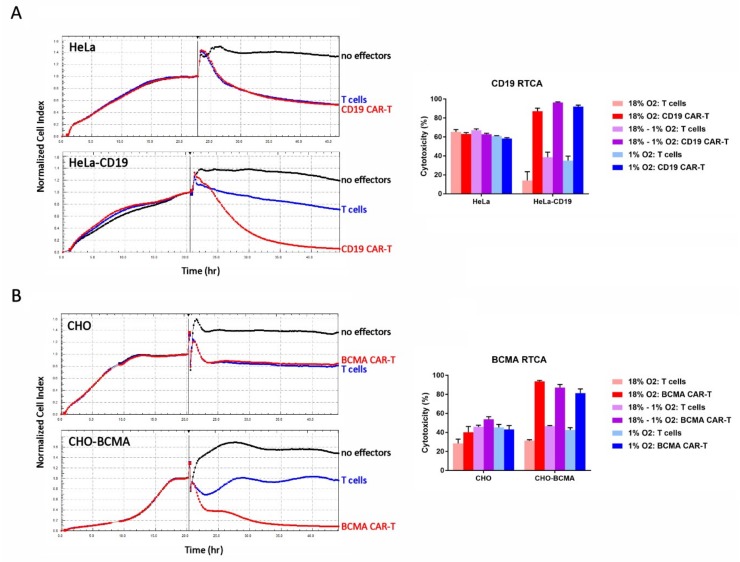
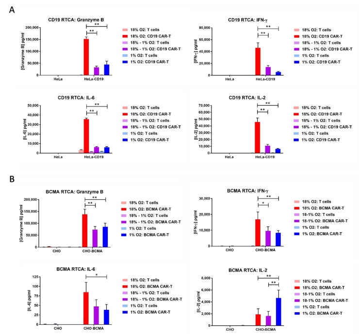
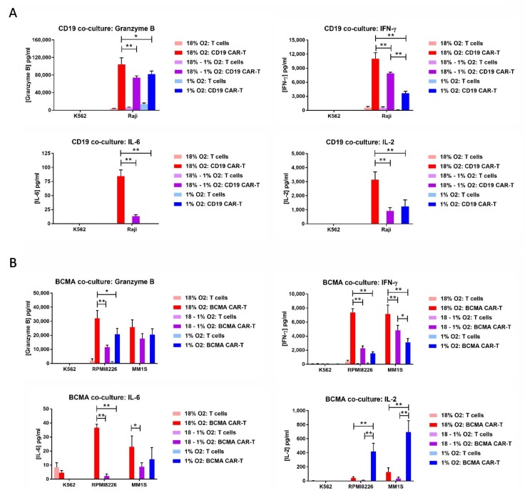
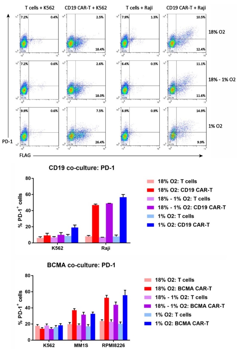
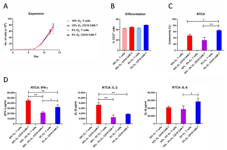
Hypoxia is a major characteristic of the solid tumor microenvironment. To understand how chimeric antigen receptor-T cells (CAR-T cells) function in hypoxic conditions, we characterized CD19-specific and BCMA-specific human CAR-T cells generated in atmospheric (18% oxygen) and hypoxic (1% oxygen) culture for expansion, differentiation status, and CD4:CD8 ratio. CAR-T cells expanded to a much lower extent in 1% oxygen than in 18% oxygen. Hypoxic CAR-T cells also had a less differentiated phenotype and a higher CD4:CD8 ratio than atmospheric CAR-T cells. CAR-T cells were then added to antigen-positive and antigen-negative tumor cell lines at the same or lower oxygen level and characterized for cytotoxicity, cytokine and granzyme B secretion, and PD-1 upregulation. Atmospheric and hypoxic CAR-T cells exhibited comparable cytolytic activity and PD-1 upregulation; however, cytokine production and granzyme B release were greatly decreased in 1% oxygen, even when the CAR-T cells were generated in atmospheric culture. Together, these data show that at solid tumor oxygen levels, CAR-T cells are impaired in expansion, differentiation and cytokine production. These effects may contribute to the inability of CAR-T cells to eradicate solid tumors seen in many patients.

Keywords: BCMA; CAR-T; CD19; hypoxia; immunotherapy; microenvironment; tumor.


Conflict of interest statement

Robert Berahovich, Xianghong Liu, Hua Zhou, Elias Tsadik, Shirley Xu, Vita Golubovskaya and Lijun Wu are employees of Promab Biotechnologies. Other authors declare no conflict of interest.

Figures

Figure 1
Figure 1
Hypoxia decreases chimeric antigen receptor-T cell (CAR-T cell) expansion. CD19 CAR-T cells (A) and B cell maturation antigen (BCMA) CAR-T cells (B), along with control T cells, were cultured in an 18% oxygen incubator for the entire 13-day expansion period (red lines), or were cultured in the 18% oxygen incubator for the first 5 days and then in a hypoxia chamber for the remaining 8 days (blue lines). Data-points represent the average and standard error of 4 separate experiments. * p = 0.02 (day 12) and ** p < 0.001 (day 13) for hypoxic vs. atmospheric CD19 CAR-T cells. ** p < 0.001 for hypoxic vs. atmospheric BCMA CAR-T cells.
Figure 2
Figure 2
Hypoxia does not affect CAR-T cell frequency. CD19 CAR-T cells (A) and BCMA CAR-T cells (B) were stained with an anti-FLAG antibody or BCMA protein, respectively. Representative flow cytometry plots showing CAR expression on the X-axis (the Y-axis is an empty channel) are on the left. Charts showing the average and standard error of 4 separate experiments are shown on the right.
Figure 3
Figure 3
Hypoxia inhibits CAR-T cell differentiation. PBMC (A), CD19 CAR-T cells (B) and BCMA CAR-T cells (C) were stained with antibodies for CD27 and CD45RO. CAR-T cells were first gated using the anti-FLAG antibody or BCMA protein. Representative flow cytometry plots showing CD27 and CD45RO expression are on the left; the CAR-T plots show only the gated CAR-T cells. Charts showing the average and standard error of 4 separate experiments are shown on the right. * p < 0.05 and ** p < 0.005.
Figure 4
Figure 4
Hypoxia increases the CD4:CD8 ratio. CD19 CAR-T cells (A) and BCMA CAR-T cells (B) were stained with antibodies for CD27 and CD45RO, along with the anti-FLAG antibody or BCMA protein. Representative flow cytometry plots showing CD27 and CD45RO expression are on the left; the CAR-T plots show only the gated CAR-T cells. Charts showing the average and standard error of 4 separate experiments are shown on the right. * p = < 0.05.
Figure 5
Figure 5
Hypoxia does not affect CAR-T cell cytotoxicity. (A) CD19 CAR-T cell RTCA assay. (B) BCMA CAR-T cell RTCA assay. Left: HeLa, HeLa-CD19, CHO, and CHO-BCMA cells were monitored overnight as they adhered to the plate and formed a monolayer. The next day, atmospheric CD19 CAR-T cells, BCMA CAR-T cells or control T cells were added to the monolayers at an E:T ratio of 10:1 (vertical bars). The cultures were monitored for approximately 24 more hours. Traces show the average of 3 wells. Right: Cytotoxicity in the RTCA assays was calculated at the end of the assays. Data-points represent the average and standard error of 4 separate experiments.
Figure 6
Figure 6
Hypoxia decreases CAR-T cell granzyme B and cytokine production. The media from the CD19 RTCA assay (A) and BCMA RTCA assay (B) was analyzed by ELISA for the levels of granzyme B, IFN-γ, IL-2 and IL-6. Data-points represent the average and standard error of 2–4 separate experiments. * p < 0.05 and ** p < 0.005.
Figure 7
Figure 7
Hypoxia decreases CAR-T cell granzyme B and cytokine production in response to tumor cells. CD19 CAR-T cells or control T cells (A) were co-cultured with CD19+ Raji cells or CD19 K562 cells. BCMA CAR-T cells or control T cells (B) were cultured with BCMA+ RPMI8226 cells, BCMA+ MM1S cells or BCMA K562 cells. The medium from the co-cultures was analyzed by ELISA for the levels of granzyme B, IFN-γ, IL-2, and IL-6. Data-points represent the average and standard error of 2-4 separate experiments. * p < 0.05 and ** p < 0.005.
Figure 8
Figure 8
Hypoxia does not affect CAR-T cell PD-1 upregulation. Top: the cells from the CD19 CAR-T cell co-cultures were analyzed by flow cytometry for FLAG staining (i.e., CD19 CAR expression) vs. PD-1 expression. Bottom: the percentages of CD19 CAR-T cells, BCMA CAR-T cells, or control T cells expressing PD-1 were plotted; data-points represent the average and standard error of 2–4 separate experiments.
Figure 9
Figure 9
CAR-T cell expansion in 5% oxygen results in greater cytotoxicity and decreased IFN-γ/IL-2 production. CD19 CAR-T cells and control T cells expanded in 18% oxygen or 5% oxygen were analyzed for cell expansion (A), differentiation (B), cytotoxicity (C), and cytokine production during the RTCA assay (D). Data points indicate averages of 2–3 replicates; * p < 0.05 and ** p < 0.005.

Similar articles

Cited by

References

    1. Maus M.V., June C.H. Making better chimeric antigen receptors for adoptive T-cell therapy. Clin. Cancer Res. 2016;22:1875–1884. doi: 10.1158/1078-0432.CCR-15-1433. - DOI - PMC - PubMed
    1. Porter D.L., Levine B.L., Kalos M., Bagg A., June C.H. Chimeric antigen receptor-modified T cells in chronic lymphoid leukemia. N. Engl. J. Med. 2011;365:725–733. doi: 10.1056/NEJMoa1103849. - DOI - PMC - PubMed
    1. Grupp S.A., Kalos M., Barrett D., Aplenc R., Porter D.L., Rheingold S.R., Teachey D.T., Chew A., Hauck B., Wright J.F., et al. Chimeric antigen receptor-modified T cells for acute lymphoid leukemia. N. Engl. J. Med. 2013;368:1509–1518. doi: 10.1056/NEJMoa1215134. - DOI - PMC - PubMed
    1. Feins S., Kong W., Williams E.F., Milone M.C., Fraietta J.A. An introduction to chimeric antigen receptor (CAR) T cell immunotherapy for human cancer. Am. J. Hematol. 2019;94:S3–S9. doi: 10.1002/ajh.25418. - DOI - PubMed
    1. Abken H. Adoptive therapy with CAR redirected T cells: The challenges in targeting solid tumors. Immunotherapy. 2015;7:535–544. doi: 10.2217/imt.15.15. - DOI - PubMed