. 2019 Apr 30;8(5):402.
doi: 10.3390/cells8050402.

Sh3bp2 Gain-Of-Function Mutation Ameliorates Lupus Phenotypes in B6.MRL- Faslpr Mice

Affiliations

Sh3bp2 Gain-Of-Function Mutation Ameliorates Lupus Phenotypes in B6.MRL- Faslpr Mice

Akiko Nagasu et al. Cells. .

Abstract

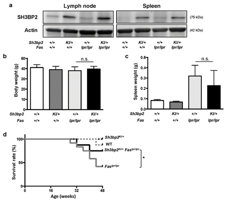
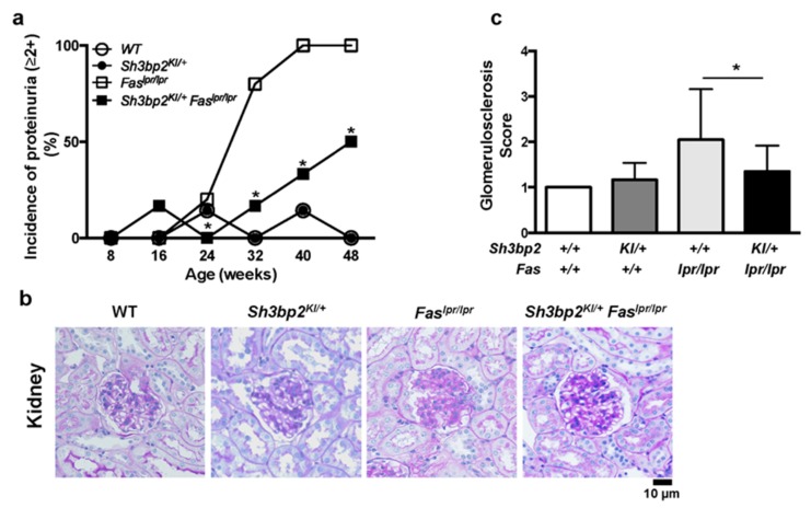
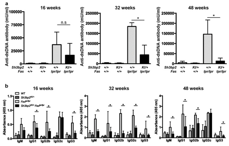
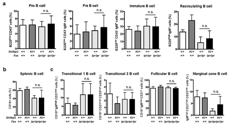
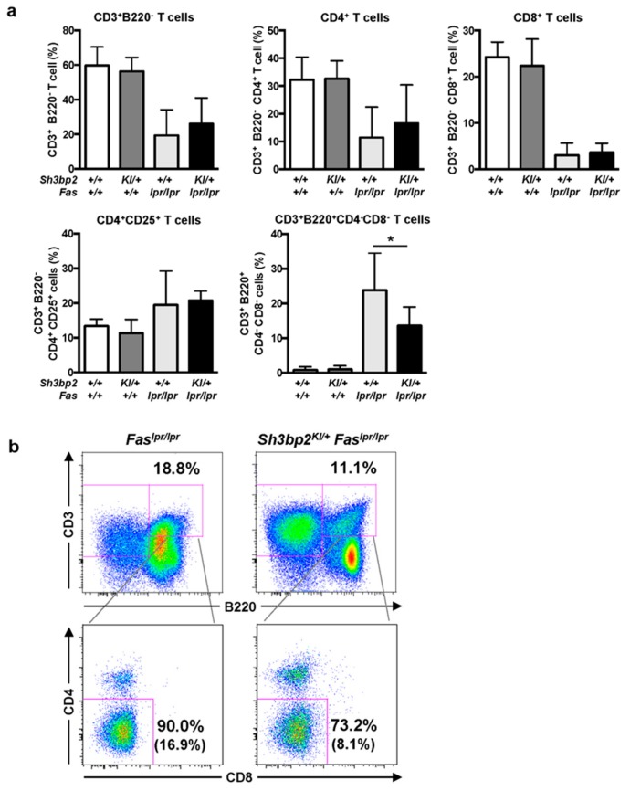
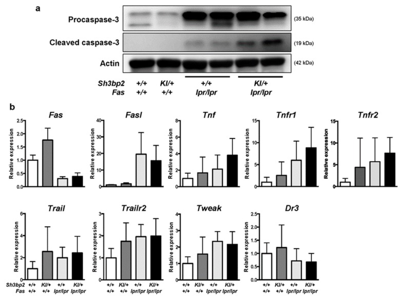
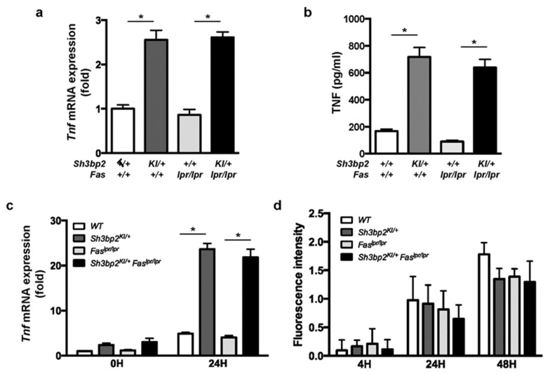
SH3 domain-binding protein 2 (SH3BP2) is an adaptor protein that is predominantly expressed in immune cells, and it regulates intracellular signaling. We had previously reported that a gain-of-function mutation in SH3BP2 exacerbates inflammation and bone loss in murine arthritis models. Here, we explored the involvement of SH3BP2 in a lupus model. Sh3bp2 gain-of-function (P416R knock-in; Sh3bp2KI/+) mice and lupus-prone B6.MRL-Faslpr mice were crossed to yield double-mutant (Sh3bp2KI/+Faslpr/lpr) mice. We monitored survival rates and proteinuria up to 48 weeks of age and assessed renal damage and serum anti-double-stranded DNA antibody levels. Additionally, we analyzed B and T cell subsets in lymphoid tissues by flow cytometry and determined the expression of apoptosis-related molecules in lymph nodes. Sh3bp2 gain-of-function mutation alleviated the poor survival rate, proteinuria, and glomerulosclerosis and significantly reduced serum anti-dsDNA antibody levels in Sh3bp2KI/+Faslpr/lpr mice. Additionally, B220+CD4-CD8- T cell population in lymph nodes was decreased in Sh3bp2KI/+Faslpr/lpr mice, which is possibly associated with the observed increase in cleaved caspase-3 and tumor necrosis factor levels. Sh3bp2 gain-of-function mutation ameliorated clinical and immunological phenotypes in lupus-prone mice. Our findings offer better insight into the unique immunopathological roles of SH3BP2 in autoimmune diseases.

Keywords: Fas; SH3 domain–binding protein 2; anti-dsDNA antibody; dendritic cells; double-negative T cells; lpr mutation; macrophages; murine lupus model; systemic lupus erythematosus; tumor necrosis factor.


Conflict of interest statement

A.N., T.M., K.K., S.T., and Y.M. received scholarship donations from Chugai Pharmaceutical Company. The funder had no role in the design of the study, collection, analyses, or interpretation of data, writing of the manuscript, or in the decision to publish the results.

Figures

Figure 1
Figure 1
Sh3bp2 gain-of-function mutation improves the survival rate of Faslpr lupus-prone mice. (a) Immunoblot analysis for SH3BP2. Protein samples were collected from lymph nodes and spleens of WT, Sh3bp2KI/+, Faslpr/lpr, and Sh3bp2KI/+Faslpr/lpr mice. Actin was used as a loading control. (bd) WT (n = 8), Sh3bp2KI/+ (n = 7), Faslpr/lpr (n = 12), and Sh3bp2KI/+Faslpr/lpr mice (n = 8) were monitored until the age of 48 weeks. At the end of the observation period, body weight (b) and spleen weight (c) were measured. (d) Survival rates of the mice. Values are presented as the mean ± SD. Note: * p < 0.05; n.s. = not significant. SH3BP2, SH3 domain-binding protein 2; WT, wild-type; KI, knock-in.
Figure 2
Figure 2
Sh3bp2 gain-of-function mutation improves renal involvement in Faslpr lupus-prone mice. (a) Incidence of proteinuria. WT (n = 7), Sh3bp2KI/+ (n = 7), Faslpr/lpr (n = 5), and Sh3bp2KI/+Faslpr/lpr mice (n = 6) were monitored until the age of 48 weeks, and levels of proteinuria were monitored every 8 weeks and graded as follows: 0, <30 mg/dL; 1+, 30–99 mg/dL; 2+, 100–299 mg/dL; 3+, >300 mg/dL. Incidence of proteinuria (≥2+) is presented. (b) Images of periodic acid-Schiff (PAS)-stained kidney sections from 48-week-old mice. Original magnification, 400×. Bar, 10 μm. (c) Renal glomerulosclerosis score. Severity of glomerulosclerosis was graded from 0 to 4, with 15 to 20 glomeruli graded per mouse. Values are presented as the mean ± SD. Note: * p < 0.05. SH3BP2, SH3 domain-binding protein 2; WT, wild-type; KI, knock-in
Figure 3
Figure 3
Sh3bp2 gain-of-function mutation suppresses aberrant anti-dsDNA antibody and immunoglobulin production in Faslpr lupus-prone mice. (a,b) Serum samples were collected from WT (n = 7), Sh3bp2KI/+ (n = 7), Faslpr/lpr (n = 5), and Sh3bp2KI/+Faslpr/lpr mice (n = 5) at 16, 32, and 48 weeks of age. Levels of anti-dsDNA antibody (a) and each immunoglobulin subclass (b) were determined by ELISA. Values are presented as the mean ± SD. * p < 0.05; n.s. = not significant. Note: dsDNA, double-stranded DNA; SH3BP2, SH3 domain-binding protein 2; WT, wild-type; KI, knock-in; ELISA, enzyme-linked immunosorbent assay.
Figure 4
Figure 4
Antibody production against thymus-independent (TI) and thymus-dependent (TD) antigens. Antibody production was comparable between WT (n = 6) and Sh3bp2KI/+ (n = 6) mice. (a) For TI antigen experiments, 8-week-old WT and Sh3bp2KI/+ mice were immunized with TNP-Ficoll. Blood samples were collected at 0-, 7-, and 14-days post-immunization, and levels of the anti-TNP antibody (IgG3) in serum (1:100 dilution) were measured by ELISA. (b) For TD antigen experiments, 8-week-old WT (n = 3) and Sh3bp2KI/+ (n = 5) mice were immunized with TNP-KLH and given a booster injection with the same dose at day 14. Blood samples were collected at 0-, 7-, 14-, and 21-days post-immunization, and levels of the anti-TNP antibody (IgG1) in serum (1:1000 dilution) were measured by ELISA. Note: SH3BP2, SH3 domain-binding protein 2; WT, wild-type; KI, knock-in; TNP, trinitrophenol; KLH, keyhole limpet hemocyanin; ELISA, enzyme-linked immunosorbent assay
Figure 5
Figure 5
B cell differentiation and maturation are unaltered in Sh3bp2KI/+Faslpr/lpr mice. (a–c) B cell subsets were analyzed in the bone marrow (a) and spleen (b,c) by flow cytometry. Cells were collected from WT (n = 7), Sh3bp2KI/+ (n = 7), Faslpr/lpr (n = 5), and Sh3bp2KI/+Faslpr/lpr mice (n = 6) at 48 weeks of age, and the suspended cells were stained with fluorochrome-labeled antibodies against IgM, IgD, CD43, and B220 for bone marrow cells (a) and IgM, IgD, CD19, CD23, and CD21/35 for splenic cells (b,c). All cells were initially gated as 7-AAD-negative single cells, followed by being gated as lymphocytes (a,b) or CD19+ cells (c). Values are presented as the mean ± SD. Note: * p < 0.05; n.s. = not significant. SH3BP2, SH3 domain-binding protein 2; WT, wild-type; KI, knock-in; 7-AAD, 7-aminoactinomycin D.
Figure 6
Figure 6
CD3+B220+CD4CD8 DNT cells are reduced in the lymph nodes of Sh3bp2KI/+Faslpr/lpr mice. (a,b) Lymph node cells were collected from WT (n = 7), Sh3bp2KI/+ (n = 7), Faslpr/lpr (n = 5), and Sh3bp2KI/+Faslpr/lpr mice (n = 6) at 48 weeks of age, and T cell subsets were stained with fluorochrome-labeled antibodies against CD3, CD4, CD8, CD25, and B220. (a) The ratio of T cell subsets was analyzed by flow cytometry. All cells were gated as 7-AAD-negative single cells, followed by being gated as lymphocytes. Values are presented as the mean ± SD; * P < 0.05; n.s. = not significant. (b) Representative flow cytometry plots of DNT cells in the lymph nodes. Flow cytometry shows a decreased proportion of DNT cells in the lymphocyte fraction of Sh3bp2KI/+Faslpr/lpr cells. The number in parentheses indicates the percentage of DNT cells in total lymphocytes. Note: DNT, double-negative T cell; SH3BP2, SH3 domain-binding protein 2; WT, wild-type; KI, knock-in; 7-AAD, 7-aminoactinomycin D.
Figure 7
Figure 7
Levels of cleaved caspase-3 are elevated in the lymph nodes of Sh3bp2KI/+Faslpr/lpr mice. (a) Images of immunoblot for procaspase-3 and cleaved caspase-3. Protein samples were collected from the lymph nodes of WT (n = 4), Sh3bp2KI/+ (n = 4), Faslpr/lpr (n = 4), and Sh3bp2KI/+Faslpr/lpr (n = 4) mice. Representative images for procaspase-3 and cleaved caspase-3 are presented. Actin was used as a loading control. (b) qPCR analysis of apoptosis-related genes. RNA samples were collected from the lymph nodes of WT (n = 7), Sh3bp2KI/+ (n = 7), Faslpr/lpr (n = 5), and Sh3bp2KI/+Faslpr/lpr (n = 6) mice at 48 weeks of age, and gene-expression levels relative to that of Hprt were determined and normalized against levels in WT samples. Values are presented as the mean ± SD. Note: SH3BP2, SH3 domain-binding protein 2; WT, wild-type; KI, knock-in; Fasl, Fas ligand; Tnf, tumor necrosis factor; Tnfr1, TNF receptor 1; Tnfr2, TNF receptor type 2; Trail, TNF-related apoptosis-inducing ligand; Trailr2, TRAIL receptor 2; Tweak, TNF-like weak inducer of apoptosis; Dr3, death receptor 3.
Figure 8
Figure 8
TNF is highly expressed in the Sh3bp2 gain-of-function mutant DCs and macrophages. (a,b) Bone marrow cells were isolated from 14- to 15-week-old WT, Sh3bp2KI/+, Faslpr/lpr, and Sh3bp2KI/+Faslpr/lpr mice and pre-cultured with GM-CSF (20 ng/mL) and IL-4 (5 ng/mL) for 8 days; resulting BMDCs were used for the experiments. (a) Tnf mRNA levels relative to that of Hprt were determined by qPCR. (b) TNF protein levels in culture supernatants. Culture supernatants were collected at the end of BMDC culture, and TNF levels were determined by ELISA. (c) Tnf mRNA expression in BMMs. Bone marrow cells were isolated from 10- to 12-week-old WT, Sh3bp2KI/+, Faslpr/lpr, and Sh3bp2KI/+Faslpr/lpr mice and stimulated with M-CSF (25 ng/mL) for 2 days, after which the yielded BMMs were treated with LPS (1 ng/mL) in the presence of M-CSF (25 ng/mL). Tnf mRNA levels relative to that of Hprt were determined by qPCR. (d) Phagocytic capacity of BMMs. Apoptotic Jurkat cells were labeled with pH-sensitive fluorescent dye and co-cultured with BMMs, followed by the measurement of fluorescence intensity derived from the engulfed apoptotic cells. Values are presented as the mean ± SD. Note: * P < 0.05. SH3BP2, SH3 domain-binding protein 2; WT, wild-type; KI, knock-in; BMDC, bone marrow-derived dendritic cell; BMM, bone marrow-derived macrophage; GM-CSF, granulocyte-macrophage colony stimulating factor; M-CSF, macrophage colony stimulating factor; IL-4, interleukin-4; LPS, lipopolysaccharide; Hprt, hypoxanthine phosphoribosyltransferase.

Similar articles

Cited by

References

    1. Tsokos G.C., Lo M.S., Costa Reis P., Sullivan K.E. New insights into the immunopathogenesis of systemic lupus erythematosus. Nat. Rev. Rheumatol. 2016;12:716–730. doi: 10.1038/nrrheum.2016.186. - DOI - PubMed
    1. Lisnevskaia L., Murphy G., Isenberg D. Systemic lupus erythematosus. Lancet. 2014;384:1878–1888. doi: 10.1016/S0140-6736(14)60128-8. - DOI - PubMed
    1. Zharkova O., Celhar T., Cravens P.D., Satterthwaite A.B., Fairhurst A.M., Davis L.S. Pathways leading to an immunological disease: Systemic lupus erythematosus. Rheumatology. 2017;56:i55–i66. doi: 10.1093/rheumatology/kew427. - DOI - PMC - PubMed
    1. Moulton V.R., Suarez-Fueyo A., Meidan E., Li H., Mizui M., Tsokos G.C. Pathogenesis of Human Systemic Lupus Erythematosus: A Cellular Perspective. Trends Mol. Med. 2017;23:615–635. doi: 10.1016/j.molmed.2017.05.006. - DOI - PMC - PubMed
    1. Furukawa F., Yoshimasu T. Animal models of spontaneous and drug-induced cutaneous lupus erythematosus. Autoimmun. Rev. 2005;4:345–350. doi: 10.1016/j.autrev.2005.01.006. - DOI - PubMed

Publication types

MeSH terms