. 2019 Apr 30;11(5):991.
doi: 10.3390/nu11050991.

Spilanthol from Traditionally Used Spilanthes acmella Enhances AMPK and Ameliorates Obesity in Mice Fed High-Fat Diet

Affiliations

Spilanthol from Traditionally Used Spilanthes acmella Enhances AMPK and Ameliorates Obesity in Mice Fed High-Fat Diet

Wen-Chung Huang et al. Nutrients. .

Abstract

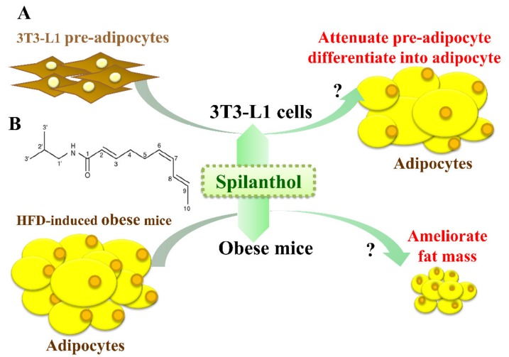
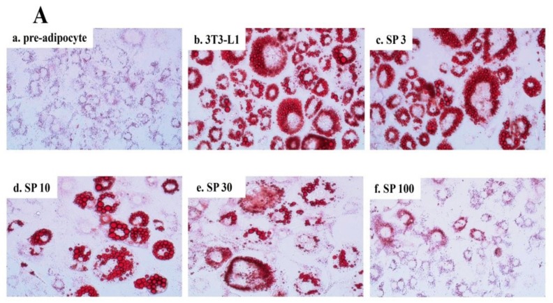
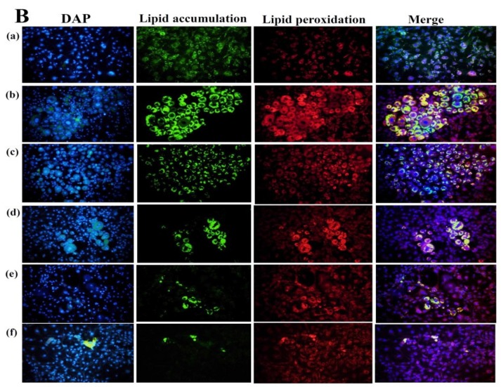
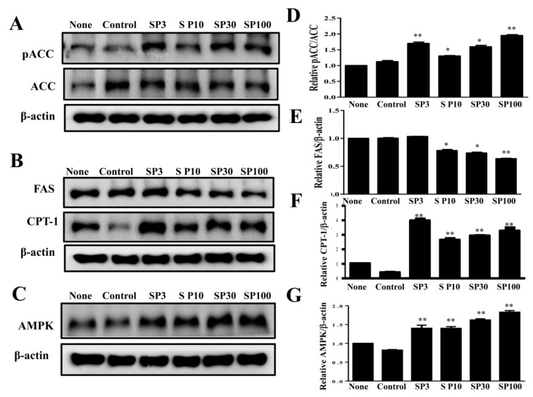
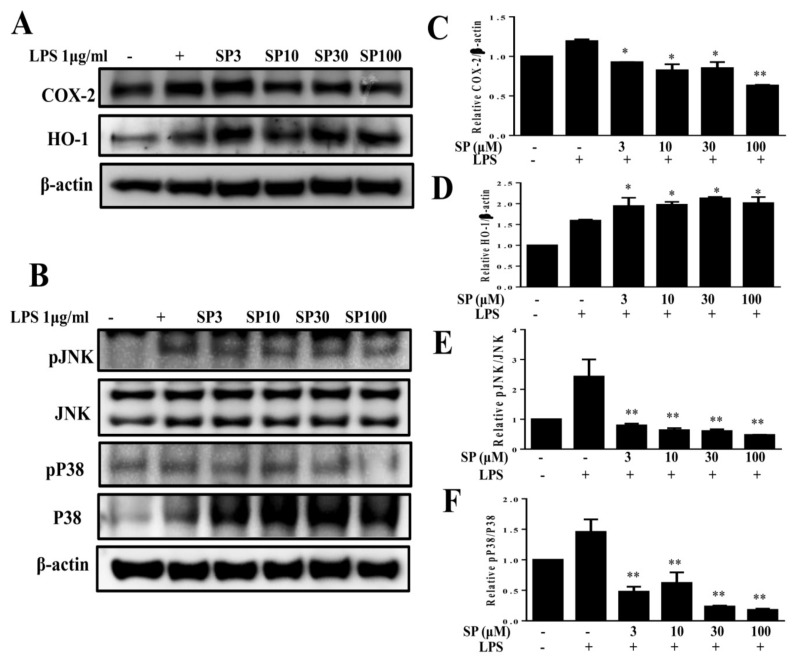
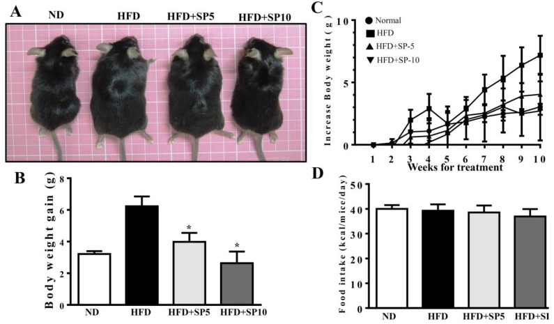
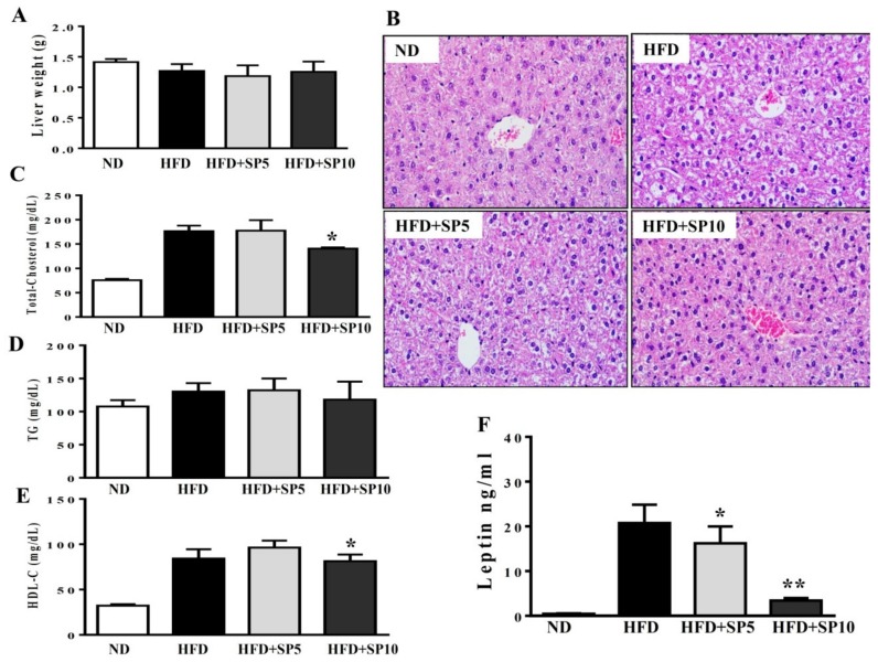
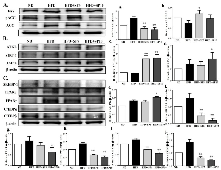
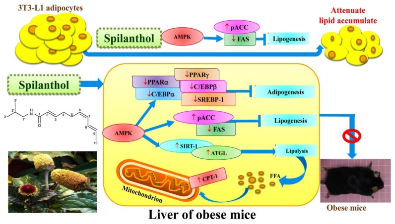
Spilanthol (SP) is a bioactive compound found in Spilanthes acmella, giving the flowers and leaves a spicy taste. Studies found that phyto-ingredients stored in spice plants act against obesity-related diseases. SP has antimicrobial, anti-inflammatory, and analgesic properties, but the effects on obesity are not yet known. We investigated the effects of SP in differentiated adipocytes (3T3-L1 cells) and mice fed a high-fat diet (HFD). SP significantly inhibited intracellular lipid accumulation and significantly reduced the expression of lipogenesis-related proteins, including acetyl-CoA carboxylase (ACC) and fatty-acid synthase (FAS). In contrast, SP increased the expression of carnitine palmitoyltransferase (CPT)1 and AMP-activated protein kinase (AMPK) in adipocytes. However, SP suppressed the levels of cyclooxygenase-2 (COX-2), phospho-p38 (pp38), and phospho-JNK (c‑Jun N-terminal kinase) (pJNK) in LPS (lipopolysaccharide)-stimulated murine pre-adipocytes. SP administered to HFD-induced obese mice via intraperitoneal injections twice a week for 10 weeks decreased body weight gain, visceral adipose tissue weight, and adipocyte size. SP inhibited lipogenic proteins FAS and ACC, and suppressed adipogenic transcription factors, enhancing lipolysis and AMPK protein expression in the liver. SP has anti-obesity effects, upregulating AMPK to attenuate lipogenic and adipogenic transcription factors.

Keywords: 3T3-L1 cells; AMPK; adipogenesis; anti-obesity; lipogenesis; spilanthol.


Conflict of interest statement

The authors declare no conflicts of interest.

Figures

Figure 1
Figure 1
Experimental abstract. (A) Study the effects of spilanthol on 3T3-L1 adipocytes and high-fat diet-induced obese mice. (B) The structure of spilanthol.
Figure 2
Figure 2
Effect of spilanthol (SP) lipid accumulation and droplets in 3T3-L1 cells. (A) 3T3-L1 pre-adipocytes were differentiated in medium containing various concentrations of SP and the differentiated adipocytes examined on day 8 by Oil Red staining: (a) 3T3-L1 pre-adipocytes; (b) 3T3-L1 adipocytes; (c) 3T3-L1 adipocytes + 3 μM SP; (d) 3T3-L1 adipocytes + 10 μM SP; (e) 3T3-L1 adipocytes + 30 μM SP; (f) 3T3-L1 adipocytes + 100 μM SP. (B) Intracellular triglyceride accumulation and lipid peroxidation measured using 4′,6-diamidino-2-phenylindole (DAPI) staining.
Figure 2
Figure 2
Effect of spilanthol (SP) lipid accumulation and droplets in 3T3-L1 cells. (A) 3T3-L1 pre-adipocytes were differentiated in medium containing various concentrations of SP and the differentiated adipocytes examined on day 8 by Oil Red staining: (a) 3T3-L1 pre-adipocytes; (b) 3T3-L1 adipocytes; (c) 3T3-L1 adipocytes + 3 μM SP; (d) 3T3-L1 adipocytes + 10 μM SP; (e) 3T3-L1 adipocytes + 30 μM SP; (f) 3T3-L1 adipocytes + 100 μM SP. (B) Intracellular triglyceride accumulation and lipid peroxidation measured using 4′,6-diamidino-2-phenylindole (DAPI) staining.
Figure 3
Figure 3
Effects of spilanthol (SP) on lipogenic and AMP-activated protein kinase (AMPK) protein expression in 3T3-L1 adipocytes. Differentiated 3T3-L1 adipocytes (104 cells/mL) were treated with 3–100 μM SP for 24 h. (A) Western blots of phosphorylated acetyl-CoA carboxylase (pACC) and ACC, (B) fatty-acid synthase (FAS) and carnitine palmitoyltransferase 1 (CPT1), and (C) AMPK protein expression. β-Actin was used as an internal control. (D) The relative protein levels of pACC/ACC, (E) FAS, (F) CPT1, and (G) AMPK. Data are presented as means ± SD (n = 3 per group); * p < 0.05, ** p < 0.01 compared to differentiated 3T3-L1 cells alone (control group). None: pre-adipocytes; control: differentiated adipocytes; SP3: differentiated adipocytes + 3 μM SP; SP10: differentiated adipocytes + 10 μM SP; SP30: differentiated adipocytes + 30 μM SP; SP100: differentiated adipocytes + 100 μM SP.
Figure 4
Figure 4
Spilanthol (SP) decreased cyclooxygenase-2 (COX-2), enhanced heme oxygenase-1 (HO-1) protein expression, and inhibited phosphorylation of mitogen-activated protein kinase (MAPK) in LPS-induced 3T3-L1 pre-adipocytes. Here, 104 cells/mL were pretreated with 3–100 μM SP for 1 h and cultured with LPS (1 μg/mL) for 24 h. (A) Western blots of COX-2 and HO-1, (B) pJNK, JNK, pP33, and P38 proteins (n = 3 per group). β-Actin was used as an internal control. (C) The relative protein levels of COX-2, (D) HO-1, (E) pJNK/JNK, and (F) pP38/P38. Data are presented as means ± SD; * p < 0.05, ** p < 0.01 compared to differentiated 3T3-L1 cells alone (control group). None: pre-adipocytes; control: differentiated adipocytes; SP3: differentiated adipocytes + 3 μM SP; SP10: differentiated adipocytes + 10 μM SP; SP30: differentiated adipocytes + 30 μM SP; SP100: differentiated adipocytes + 100 μM SP.
Figure 5
Figure 5
Spilanthol (SP) ameliorates adiposity and decreases body weight gain in high-fat diet (HFD)-induced obese mice. (A) Representative images of the whole body. (B) Body weight (BW) and (C) body weight gain. (D) Food intake (kcal/mice/day). C57BL/6 mice were fed a normal diet (ND) or high-fat diet (HFD) with or without a low dose of SP (5 mg spilanthol/kg BW; SP5) or high dose of SP (10 mg spilanthol/kg BW; SP10) for eight weeks. The data are presented as means ± SD, n = 8; * p < 0.05 compared to HFD-fed mice alone.
Figure 6
Figure 6
Spilanthol (SP) reduces HFD-induced visceral adipocyte tissue weight and adipocyte size. (A) Representative image of epididymal adipose tissue. (B) Epididymal adipose tissue weight. (C) Epididymal adipocyte size. (D) Histological sections of epididymal adipose tissues from mice (400×). (E) Representative image of inguinal adipose tissue. (F) Inguinal adipose tissue weight. (G) Inguinal adipocyte size. (H) Histological sections of inguinal adipose tissue (400×). C57BL/6 mice were fed a normal diet (ND) or high-fat diet (HFD) with or without a low dose of SP (5 mg spilanthol/kg BW; SP5) or high dose of SP (10 mg spilanthol/kg BW; SP10) for eight weeks. The data are presented as means ± SD, n = 8; * p < 0.05, ** p < 0.01 compared to HFD-fed mice alone.
Figure 7
Figure 7
Spilanthol (SP) ameliorates hepatic steatosis, decreases serum leptin levels, and improves serum biochemical parameters in HFD-induced obese mice. (A) Final liver weight. (B) Hematoxylin and eosin (H&E)-stained liver tissue. (C) Serum total cholesterol level. (D) Serum triglyceride level. (E) Serum high-density lipoprotein cholesterol (HDL-C) level. (F) Serum leptin level. C57BL/6 mice were fed a normal diet (ND) or high-fat diet (HFD) with or without a low dose of SP (5 mg spilanthol/kg BW; SP5) or high dose of SP (10 mg spilanthol/kg BW; SP10) for eight weeks. The data are presented as means ± SD, n = 8; * p < 0.05, ** p < 0.01 compared to HFD-fed mice alone.
Figure 8
Figure 8
Treatment with spilanthol (SP) downregulates the lipogenesis and adipogenesis-related transcription factors, and upregulates lipolysis-related and AMPK pathway protein expression in the livers of HFD-fed mice. (A) Western blots of lipogenesis-related proteins FAS and pACC, (B) lipolysis protein triglyceride lipase (ATGL), sirtuin 1 (SIRT1), and AMPK, and (C) adipogenic transcription factors sterol regulatory element-binding protein (SREBP-1), peroxisome proliferator-activated receptor (PPAR)α, PPARγ, CCAAT/enhancer-binding protein (C/EBP)α, and C/EBPβ. β-Actin was used as an internal control. The relative protein levels of FAS (a), pACC (b), ATGL (c), SIRT1 (d), AMPK (e), SREBP-1 (f), PPARα (g), PPARγ (h), C/EBPα (i), and C/EBPβ (j) in the liver are also given. Data are presented as means ± SD (n = 8); * p < 0.05, ** p < 0.01 compared to HFD-induced obese mice.
Figure 9
Figure 9
Model explaining the mechanism underlying the anti-obesity effects of spilanthol (SP). SP increases lipolysis-related proteins, suppresses the protein expression of adipogenesis-related transcript factors, and reduces lipogenic proteins via activation of AMPK in the liver of obese mice. SP also inhibits lipogenesis to reduce ACC and FAS expression in 3T3-L1 adipocytes. SP is a potential anti-obesity compound that could ameliorate liver lipid accumulation in obese mice.

References

    1. Seong J., Kang J.Y., Sun J.S., Kim K.W. Hypothalamic inflammation and obesity: A mechanistic review. Arch. Pharm. Res. 2019:5. doi: 10.1007/s12272-019-01138-9. - DOI - PubMed
    1. Ellulu M.S., Patimah I., Khaza’ai H., Rahmat A., Abed Y. Obesity and inflammation: The linking mechanism and the complications. Arch. Med. Sci. 2017;13:851–863. doi: 10.5114/aoms.2016.58928. - DOI - PMC - PubMed
    1. Hirabara S.M., Gorjão R., Vinolo M.A., Rodrigues A.C., Nachbar R.T., Curi R. Molecular targets related to inflammation and insulin resistance and potential interventions. J. Biomed. Biotechnol. 2012:379024. doi: 10.1155/2012/379024. - DOI - PMC - PubMed
    1. Tateya S., Kim F., Tamori Y. Recent advances in obesity-induced inflammation and insulin resistance. Front. Endocrinol. 2013;4:93. doi: 10.3389/fendo.2013.00093. - DOI - PMC - PubMed
    1. Seth S., Martin B.S., Qasim A., Muredach P., Reilly M.B. Leptin resistance: A Possible interface of inflammation and metabolism in obesity-related cardiovascular disease. J. Am. Coll. Cardiol. 2008;7:1201–1210. - PMC - PubMed