. 2019 May 3;18(1):159.
doi: 10.1186/s12936-019-2790-2.

Comparing sequence and structure of falcipains and human homologs at prodomain and catalytic active site for malarial peptide based inhibitor design

Affiliations

Comparing sequence and structure of falcipains and human homologs at prodomain and catalytic active site for malarial peptide based inhibitor design

Thommas Mutemi Musyoka et al. Malar J. .

Abstract

Background: Falcipains are major cysteine proteases of Plasmodium falciparum involved in haemoglobin degradation and remain attractive anti-malarial drug targets. Several inhibitors against these proteases have been identified, yet none of them has been approved for malaria treatment. Other Plasmodium species also possess highly homologous proteins to falcipains. For selective therapeutic targeting, identification of sequence and structure differences with homologous human cathepsins is necessary. The substrate processing activity of these proteins is tightly controlled via a prodomain segment occluding the active site which is chopped under low pH conditions exposing the catalytic site. Current work characterizes these proteases to identify residues mediating the prodomain regulatory function for the design of peptide based anti-malarial inhibitors.

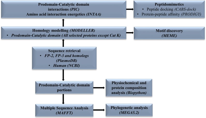
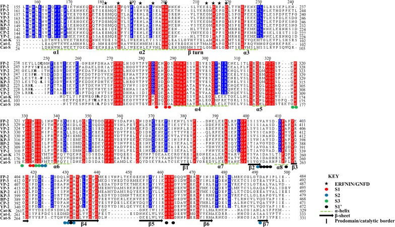
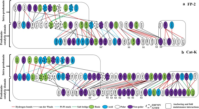
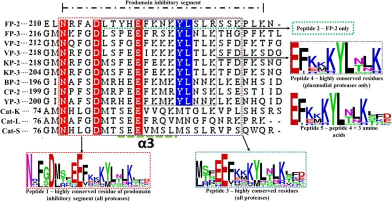
Methods: Sequence and structure variations between prodomain regions of plasmodial proteins and human cathepsins were determined using in silico approaches. Additionally, evolutionary clustering of these proteins was evaluated using phylogenetic analysis. High quality partial zymogen protein structures were modelled using homology modelling and residue interaction analysis performed between the prodomain segment and mature domain to identify key interacting residues between these two domains. The resulting information was used to determine short peptide sequences which could mimic the inherent regulatory function of the prodomain regions. Through flexible docking, the binding affinity of proposed peptides on the proteins studied was evaluated.

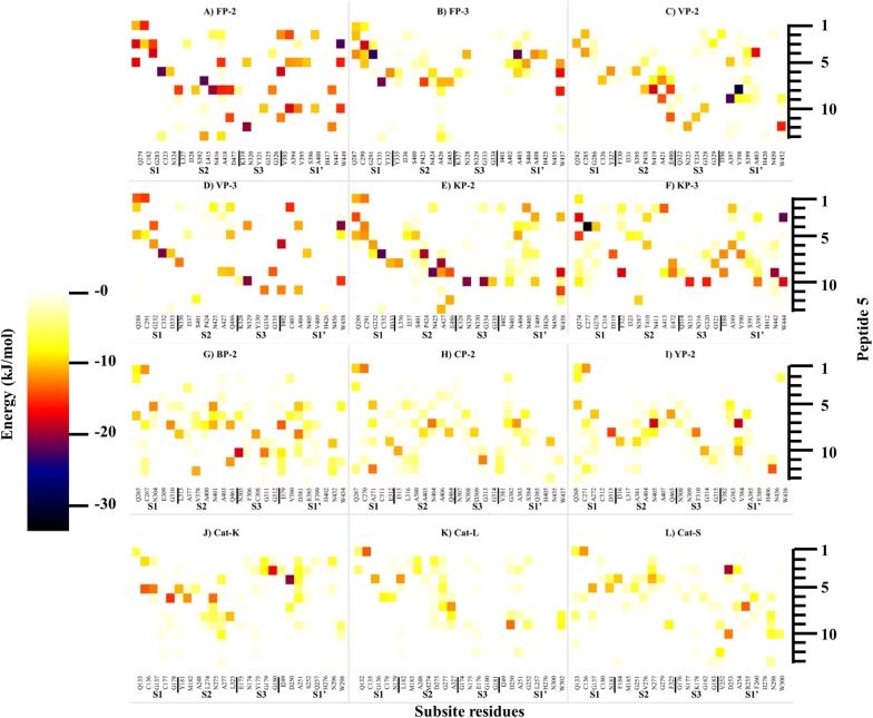
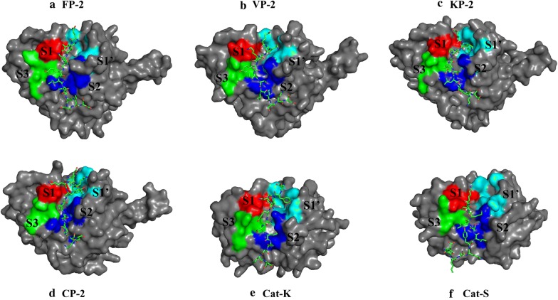
Results: Sequence, evolutionary and motif analyses showed important differences between plasmodial and human proteins. Residue interaction analysis identified important residues crucial for maintaining prodomain integrity across the different proteins as well as the pro-segment responsible for inhibitory mechanism. Binding affinity of suggested peptides was highly dependent on their residue composition and length.

Conclusions: Despite the conserved structural and catalytic mechanism between human cathepsins and plasmodial proteases, current work revealed significant differences between the two protein groups which may provide valuable information for selective anti-malarial inhibitor development. Part of this study aimed to design peptide inhibitors based on endogenous inhibitory portions of protease prodomains as a novel aspect. Even though peptide inhibitors may not be practical solutions to malaria at this stage, the approach followed and results offer a promising means to find new malarial inhibitors.

Keywords: Binding affinity; Cysteine protease; Falcipain; Homology modelling; Prodomain inhibitory segment; Zymogen.


Conflict of interest statement

The authors declare that they have no competing interests.

Figures

Fig. 1
Fig. 1
Clan CA cysteine protease zymogen prodomain-catalytic domain interaction modes. Surface representation of a human Cat-K and b FP-2. c FP-2 prodomain structural elements (pink; in cartoon representation) interacting with the S1 (red), S2 (blue), S3 (green) and S1′ (cyan) subsites of the catalytic domain
Fig. 2
Fig. 2
A graphical workflow of the methods and tools (in brackets) used in sequence and structural analysis of FP-2, FP-3 and their homologs
Fig. 3
Fig. 3
Homology models of different plasmodial proteases and human Cat-L together with the templates used in homology modelling. Colour code ranges from blue (accurate modelling) to red (poorly modelled regions)
Fig. 4
Fig. 4
Structural-based multiple sequence alignment of FP-2, FP-3 and homologs prodomain-catalytic domains. Actual residue numbering per protein is given on the side, and the top numbering is based on partial zymogen alignment. The papain family characteristic prodomain ERFNIN and GNFD motif residues are indicated with an asterisk. Bold short lines depict the prodomain-catalytic domain border. Dashed green lines indicate the position of α-helix and arrows β-sheet structural elements. Fully conserved residues in all the proteins are marked with red while residues only conserved in plasmodial proteases with blue. Position of subsite residues is shown with filled circles (Red = S1, Blue = S2, Green = S3 and S1′ = black)
Fig. 5
Fig. 5
A phylogenetic tree of plasmodial and human FP-3, and FP-3 homologs prodomain-catalytic protein sequences using MEGA5.2.2. The evolutionary history was inferred by using the Maximum Likelihood method based on the Whelan and Goldman model (WAG) model with a γ discrete distribution (+G) parameter of 2.4 and an evolutionary invariable ([+I]) of 0.1. All positions with gaps were completely removed (100% deletion) and bootstrap value set at 1000. The scale bar represents the number of amino acid substitutions per site. Toxoplasma gondii CAT-L is used as the outgroup
Fig. 6
Fig. 6
Motif analysis of plasmodial proteases and human cathepsins partial zymogen domains. a A heat map showing the distribution, level of conservation and information of different motifs found in plasmodial and human proteases studied. A cartoon presentation showing the location of all motifs within the prodomain-catalytic structural fold. Labelled in green boxes are motifs present in both (b) human cathepsins and (c) plasmodial proteases
Fig. 7
Fig. 7
Intra-prodomain and prodomain-catalytic residue interaction network in a FP-2 and b Cat-K. For each protein full length residue numbering is used. Enclosed in black are residue interactions involved in anchoring the prodomain onto the catalytic domain with the rest being those involved in mediating the inhibitory effect. Shown in lines are the different interaction types between the prodomain and catalytic domains
Fig. 8
Fig. 8
A heatmap for residue interaction energies between prodomain inhibitory segment and the catalytic subsite residues per protein. The inhibitory segment starts from the conserved Asn residue in the GNFD motif (Fig. 4)
Fig. 9
Fig. 9
Sequence alignment of the prodomain inhibitory segment for the plasmodial and human cathepsin proteases studied. Marked sequence sections indicate the portions used to design different oligopeptides for docking studies and their conservation as determined by WebLogo server. Actual residue numbering per protein is given on the side
Fig. 10
Fig. 10
A heatmap for residue interaction energies between peptide 5 and the catalytic subsite residues per protein
Fig. 11
Fig. 11
Peptide 5 binding mode with catalytic domain of various proteins (Red = S1, Blue = S2, Green = S3 and Cyan = S1′)

Similar articles

Cited by

References

    1. WHO. World malaria report 2017. Geneva: World Health Organization; 2017. http://apps.who.int/iris/bitstream/handle/10665/259492/9789241565523-eng.... Accessed 8 May 2018.
    1. Bass C, Jones CM. Mosquitoes boost body armor to resist insecticide attack. Proc Natl Acad Sci USA. 2016;113:9145–9147. doi: 10.1073/pnas.1610992113. - DOI - PMC - PubMed
    1. Hemingway J, Pickett J, Ranson H, Magill A, Simard F, Fornadel C, et al. Averting a malaria disaster: will insecticide resistance derail malaria control? Lancet. 2016;387:1785–1788. doi: 10.1016/S0140-6736(15)00417-1. - DOI - PMC - PubMed
    1. Alout H, Yameogo B, Djogbénou LS, Chandre F, Dabiré RK, Corbel V, et al. Interplay between Plasmodium infection and resistance to insecticides in vector mosquitoes. J Infect Dis. 2014;210:1464–1470. doi: 10.1093/infdis/jiu276. - DOI - PubMed
    1. Tun KM, Imwong M, Lwin KM, Win AA, Hlaing TM, Hlaing T, et al. Spread of artemisinin-resistant Plasmodium falciparum in Myanmar: a cross-sectional survey of the K13 molecular marker. Lancet Infect Dis. 2015;15:415–421. doi: 10.1016/S1473-3099(15)70032-0. - DOI - PMC - PubMed

LinkOut - more resources