. 2019 Jun 20;74(6):1215-1226.e4.
doi: 10.1016/j.molcel.2019.04.005. Epub 2019 Apr 30.

PD-L1 (B7-H1) Competes with the RNA Exosome to Regulate the DNA Damage Response and Can Be Targeted to Sensitize to Radiation or Chemotherapy

Affiliations

PD-L1 (B7-H1) Competes with the RNA Exosome to Regulate the DNA Damage Response and Can Be Targeted to Sensitize to Radiation or Chemotherapy

Xinyi Tu et al. Mol Cell. .

Abstract

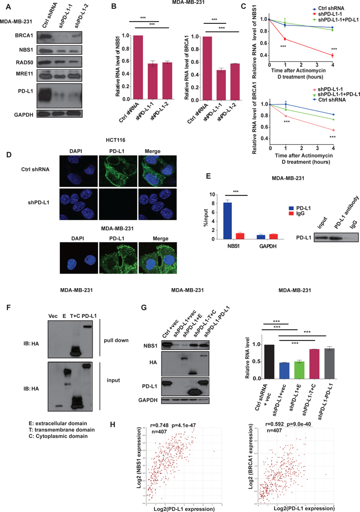
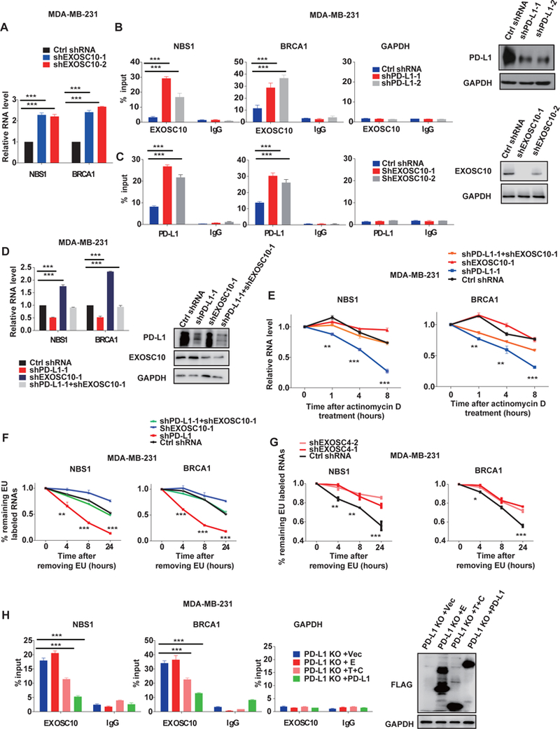
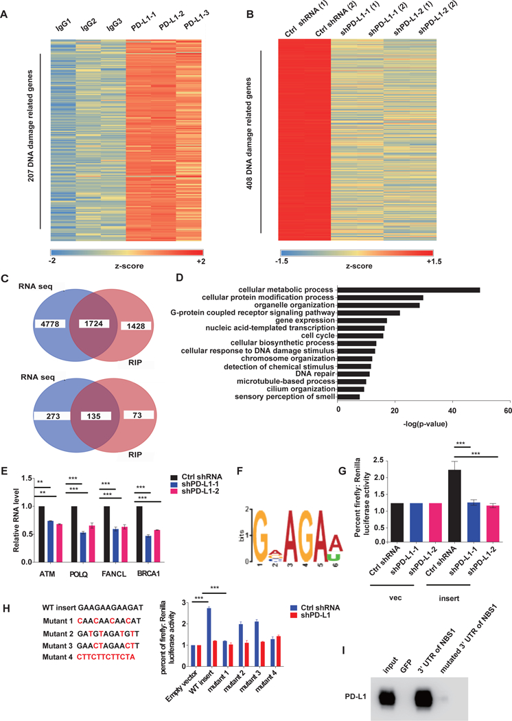
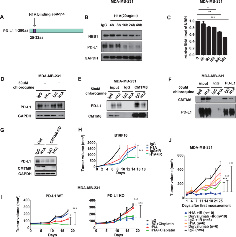
Programmed death ligand 1 (PD-L1, also called B7-H1) is an immune checkpoint protein that inhibits immune function through its binding of the programmed cell death protein 1 (PD-1) receptor. Clinically approved antibodies block extracellular PD-1 and PD-L1 binding, yet the role of intracellular PD-L1 in cancer remains poorly understood. Here, we discovered that intracellular PD-L1 acts as an RNA binding protein that regulates the mRNA stability of NBS1, BRCA1, and other DNA damage-related genes. Through competition with the RNA exosome, intracellular PD-L1 protects targeted RNAs from degradation, thereby increasing cellular resistance to DNA damage. RNA immunoprecipitation and RNA-seq experiments demonstrated that PD-L1 regulates RNA stability genome-wide. Furthermore, we developed a PD-L1 antibody, H1A, which abrogates the interaction of PD-L1 with CMTM6, thereby promoting PD-L1 degradation. Intracellular PD-L1 may be a potential therapeutic target to enhance the efficacy of radiotherapy and chemotherapy in cancer through the inhibition of DNA damage response and repair.

Keywords: CMTM6; DNA repair; PD-L1; PD-L1 destabilization; RNA binding; RNA exosome; anti-B7-H1 antibody; chemotherapy; immunotherapy; radiotherapy.


Figures

Figure. 1,
Figure. 1,. PD-L1 is required for the DNA damage response
A-B, HCT116 cells (A) or MDA-MB-231 cells (B) transfected with the indicated constructs were treated with either ionizing radiation (IR) or cisplatin at the indicated doses and colony formation was assessed (±s.e.m., n=3). (*p<0.05, **p<0.01, ***p<0.001) C, HeLa cells transfected with PD-L1 overexpression or control vectors were treated with either IR or cisplatin and colony formation was assessed (±s.e.m., n=3). (*p<0.05, **p<0.01, ***p<0.001) D, Patient-derived breast cancer cells were isolated and infected with control or PD-L1 targeted shRNA lentivirus. Cells were seeded and treated with the indicated doses of irradiation and the viability of cell was assessed cell survival (±s.e.m., n=3). (*p<0.05, **p<0.01, ***p<0.001 ) E, Control and PD-L1 knockdown U2OS cells were treated with 2 Gy and the formation and resolution of γH2AX foci were assessed using immunofluorescence. Representative images are shown with data quantification in Supplementary Figure 1B. F, Wild-type (+/+, n=16), PD-L1 heterozygous (+/−, n=24) and PD-L1 knockout (−/−, n=16) BALB/C mice were exposed to whole body irradiation (7 Gy) and survival was assessed. Comparisons between groups were made using the Log-rank test.
Figure. 2,
Figure. 2,. PD-L1 binds and stabilizes NBS1 and BRCA1 mRNA.
A, Western blot analysis of BRCA1, NBS1, RAD50, MRE11 and PD-L1 in MDA-MB-231 cells infected with lentiviruses encoding control shRNA or two different shRNAs targeting PD-L1. GAPDH was used as a loading control. B, Quantification of NBS1 and BRCA1 mRNA levels in MDA-MB-231 cells infected with lentiviruses encoding indicated shRNAs using quantitative real-time (qRT)-PCR (±s.e.m., n=3). GAPDH was used for normalization. (*p<0.05, **p<0.01, ***p<0.001) C, Control MDA-MB-231 cells, PD-L1 knockdown, and PD-L1 knockdown cells with PD-L1 re-expressed were treated with the transcription inhibitor actinomycin D (5 ug/ml). NBS1 and BRCA1 mRNA levels were quantified using qRT-PCR (±s.e.m., n=3). GAPDH was used for normalization. (*p<0.05, **p<0.01, ***p<0.001). D, Representative immunofluorescence images of the subcellular localization of PD-L1 in HCT116 cells and MDA-MB-231. PD-L1 was labeled with green fluorescence and the nucleus was labeled with DAPI. E, RNA immunoprecipitation (RIP) assay demonstrating significant enrichment of NBS1 mRNA by PD-L1 antibody compared to the negative control IgG. The result is shown as the percentage of input (±s.e.m., n=3). (*p<0.05, **p<0.01, ***p<0.001). F, RNA pull down in MDA-MB-231 cells expressing different truncations of PD-L1 (vector control, extracellular domain [E], transmembrane plus cytoplasmic domain [T+C], and full length PD-L1) using in vitro biotin labeled 3’ NBS1 RNA. G, Following PD-L1 knockdown by shRNA in MDA-MB231 cells, various truncations of PD-L1 were overexpressed and the level of NBS1 protein (left), and NBS1 mRNA (right), was assessed using immunoblot or qRT-PCR, respectively. GAPDH was using as loading controls. For qRT-PCR the result is shown as the percentage of input (±s.e.m., n=3). (*p<0.05, **p<0.01, ***p<0.001). H, The correlation of PD-L1 expression with NBS1 and BRCA1 was assessed using blood samples data from 407 healthy donors in The Cancer Genome Atlas (TCGA) database. The Pearson correlation coefficient (r) is shown with the associated p value.
Figure. 3,
Figure. 3,. PD-L1 protects NBS1 and BRCA1 mRNA from degradation by the RNA exosome
A, qRT-PCR analysis demonstrating NBS1 and BRCA1 mRNA levels following knockdown of EXOSC10 in MDA-MB-231 cells with two separate shRNA. GAPDH was used for normalization. (*p<0.05, **p<0.01, ***p<0.001). B, RIP assay in MDA-MB-231 cells demonstrating that knockdown of PD-L1 increases the binding affinity of EXOSC10 with NBS1 and BRCA1 mRNA. The result is shown as the percentage of input (±s.e.m., n=3). (*p<0.05, **p<0.01, ***p<0.001). C, RIP assay in MDA-MB-231 cells demonstrating that knockdown of EXOSC10 increases the binding affinity of PD-L1 with NBS1 and BRCA1 mRNA. The result is shown as the percentage of input (±s.e.m., n=3). (*p<0.05, **p<0.01, ***p<0.001). D, qRT-PCR analysis of NBS1 and BRCA1 mRNA levels in MDA-MB-231 cells infected with the indicated shRNAs (±s.e.m., n=3). GAPDH was used for normalization. (*p<0.05, **p<0.01, ***p<0.001). E, MDA-MB-231 cells infected with the indicated shRNAs were treated with the transcription inhibitor actinomycin D (5 ug/ml) and NBS1 (left) and BRCA1 (right) mRNA stability was assessed. (*p<0.05, **p<0.01, ***p<0.001). F and G, 5-Ethynyluridine (EU) labeling assay to determine NBS1 and BRCA1 mRNA stability under PD-L1 or EXOSC10 knockdown (F) and EXOSC4 knockdown (G) conditions. Cells were first treated with EU (0.1mM) for 24 hours, and the incorporation ratio of EU was determined. Then EU was removed and cells were cultured in fresh media for another 0, 4, 8, or 24 hours. The stability of BRCA1 and NBS1 mRNAs was determined as % of labeled mRNA after removal of EU divided by % of labeled mRNA before removal. H, RIP assay of PD-L1 knockout MDA-MB-231 cells stably transfected with indicated Flag-tagged PD-L1 truncations. The binding affinity of EXOSC10 with NBS1 (left) and BRCA1 (middle) mRNA was evaluated. GAPDH control is also displayed (right). The result is shown as the percentage of input (±s.e.m., n=3). (*p<0.05, **p<0.01, ***p<0.001).
Figure. 4,
Figure. 4,. PD-L1 regulates RNA stability genome-wide
A, Cross-linked RIP was performed in MDA-MB-231 cells using PD-L1 antibody or control IgG followed by RNA sequencing on the extracted RNAs. Heatmap of DNA damage related genes enriched by PD-L1 through RIP-seq is shown (three independent replicates). The map was plotted according to the z-score of log (number of peaks). B, MDA-MB-231 cells were depleted of PD-L1 using control or two separate shRNAs and RNA sequencing was performed. Heatmap of DNA damage genes enriched by PD-L1 is shown. The map was plotted according to the z-score of log (normalized counts). C, Venny diagram of overlapping genes from the RIP-seq and RNA-seq analyses in A and B. The RNAs that were found to interact with PD-L1 through RIP-seq in A overlapped with the downregulated RNAs caused by PD-L1 knockdown in B. D, Gene Ontology (GO) analysis of RIP-seq and RNAseq data. RNAs that interacted with PD-L1 that were also downregulated by PD-L1 knockdown were significantly enriched in multiple important biological pathways including DNA damage response and repair related pathways. The data is shown as –log (p-value). E, qRT-PCR analysis of MDA-MB-231 cells infected with lentiviruses encoding indicated shRNAs to quantify several of the DNA damage response and repair related genes enriched by both RIP-Seq and RNA-Seq data (±s.e.m., n=3). GAPDH was used for normalization. (*p<0.05, **p<0.01, ***p<0.001). F, The top PD-L1 binding RNA motif identified using MEME ChIP software. G and H, The dual luciferase reporter assay using pmirGLO vector. Two copies of the candidate RNA motif (GAAGAAGAAGAT) and mutant sequences were inserted into the 3’UTR of the firefly luciferase gene, and then both empty vector and the vector with insert were transfected into control and PD-L1 knockdown cells. The signal of firefly luciferase was measured. Renilla luciferase was used as an internal control (±s.e.m., n=6, two tails t-test). (*p<0.05, **p<0.01, ***p<0.001). I, RNA pull down assay in MDA-MB-231 lysate using either the 3’ UTR of NBS1 or the 3’ UTR of NBS1 with binding motif mutations. GFP RNA was used as a negative control.
Figure. 5
Figure. 5. The anti-PD-L1 antibody, H1A, destabilizes PD-L1 and sensitizes cancer to radio-therapy
A, Diagram of the H1A binding epitope on PD-L1 (20–32 aa). B-C, MDA-MB-231 cells were plated one day before treatment with either H1A (20 ug/ml) or control IgG. Cells were lysed at the indicated time points and NBS1 and PD-L1 expression was assessed using immunoblot (B) or qRT-PCR (C). GAPDH was used as a loading control. (*p<0.05, **p<0.01, ***p<0.001 ) D, MDA-MB-231 cells were plated one day before either H1A (20 ug/ml) or control IgG treatment, with or without the lysosome inhibitor, chloroquine, and expression of PD-L1 was assessed using immunoblot. GAPDH was used as a loading control. E-F, MDA-MB-231 cells were treated with H1A (20 ug/ml) and chloroquine (50 uM). 16 hours later, cells were harvested and immunoprecipitation was performed using CMTM6 (E) or PD-L1 (F) antibodies and CMTM6 and PD-L1 was detected using immunoblot. G, Control and CMTM6 knockout cells were treated either IgG or H1A (20ug/ml) antibody and PD-L1 level was determined by Western blot. H, human PD-L1 expressing murine melanoma B16F10 cells with endogenous PD-L1 knocked out were subcutaneously injected into the flank of C57BL/6 mice. Animals were randomized into four groups (n=5) and treated with either IgG, H1A, IgG + IR or H1A + IR. Bars represent SEM. The p value for the primary comparison of IgG + IR vs H1A + IR was calculated using a t test. (*p<0.05, **p<0.01, ***p<0.001). I, Wild-type (WT, left) or PD-L1 knock out (KO, right) MDA-MB-231 cells were subcutaneously injected into the flank of NOD-SCID mice. Animals were randomized into four groups (n=5) and treated with either control IgG, H1A, IgG + cisplatin or H1a + cisplatin. Bars represent SEM. The p value for the primary comparison of IgG + cisplatin vs H1A + cisplatin was calculated using a t test. (*p<0.05, **p<0.01, ***p<0.001 ) J, MDA-MB-231 cells were subcutaneously injected into the flank of NOD-SCID mice and mice were randomized and treated with either IgG, H1A, Durvalumab, IgG + IR, H1A + IR or Durvalumab + IR. Bars represent SEM. The p value for the comparisons of H1A + IR vs durvalumab + IR, H1A+ IR vs IgG + IR, and H1A + IR vs IgG are shown and were calculated using a t test.(*p<0.05, **p<0.01, ***p<0.001 )

References

    1. Antonia SJ, Villegas A, Daniel D, Vicente D, Murakami S, Hui R, Yokoi T, Chiappori A, Lee KH, de Wit M, et al. (2017). Durvalumab after Chemoradiotherapy in Stage III Non-Small-Cell Lung Cancer. N Engl J Med 377, 1919–1929. - PubMed
    1. Burr ML, Sparbier CE, Chan YC, Williamson JC, Woods K, Beavis PA, Lam EYN, Henderson MA, Bell CC, Stolzenburg S, et al. (2017). CMTM6 maintains the expression of PD-L1 and regulates anti-tumour immunity. Nature 549, 101–105. - PMC - PubMed
    1. Chen DS, Irving BA, and Hodi FS (2012). Molecular pathways: next-generation immunotherapy--inhibiting programmed death-ligand 1 and programmed death-1. Clin Cancer Res 18, 6580–6587. - PubMed
    1. Chen L, and Han X (2015). Anti-PD-1/PD-L1 therapy of human cancer: past, present, and future. J Clin Invest 125, 3384–3391. - PMC - PubMed
    1. Chen N, Fang W, Zhan J, Hong S, Tang Y, Kang S, Zhang Y, He X, Zhou T, Qin T, et al. (2015). Upregulation of PD-L1 by EGFR Activation Mediates the Immune Escape in EGFR-Driven NSCLC: Implication for Optional Immune Targeted Therapy for NSCLC Patients with EGFR Mutation. J Thorac Oncol 10, 910–923. - PubMed

Publication types

MeSH terms