. 2019 May 3;4(35):eaax0416.
doi: 10.1126/sciimmunol.aax0416.

Stromal cells maintain immune cell homeostasis in adipose tissue via production of interleukin-33

Affiliations

Stromal cells maintain immune cell homeostasis in adipose tissue via production of interleukin-33

T Mahlakõiv et al. Sci Immunol. .

Abstract

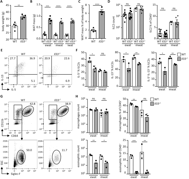
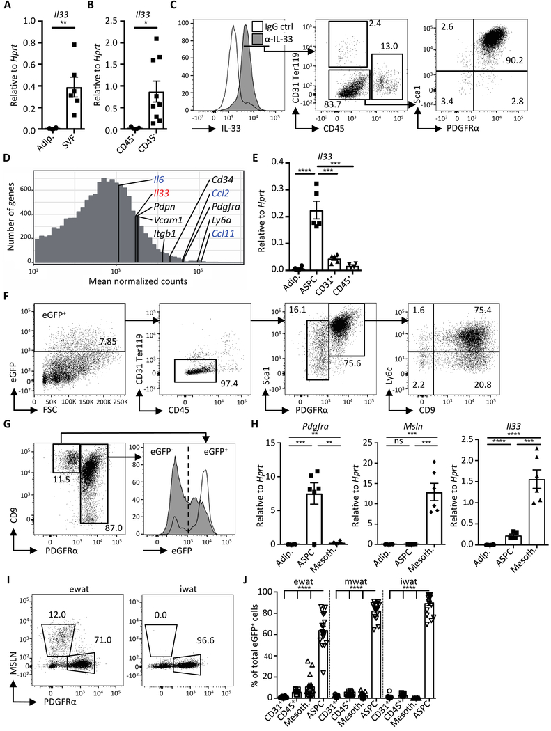
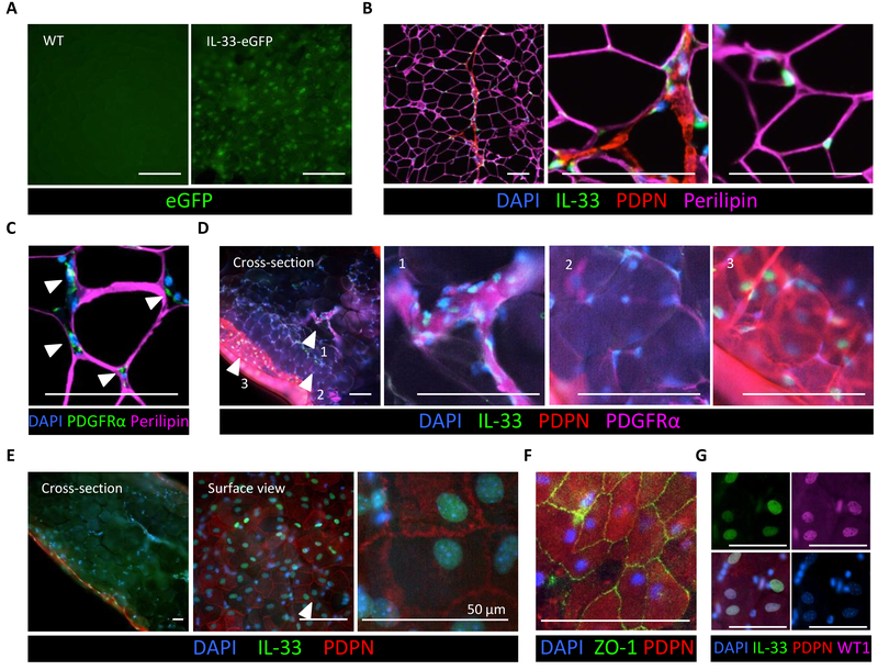
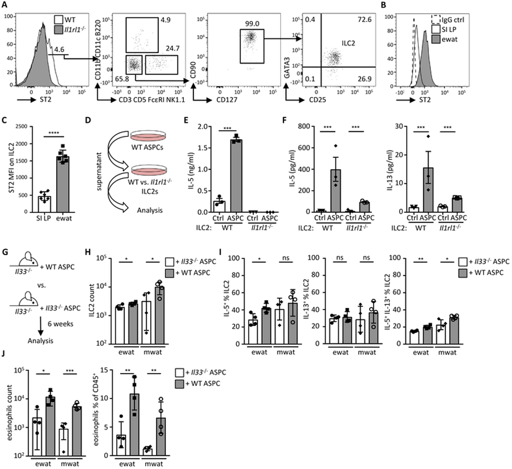
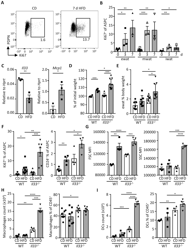
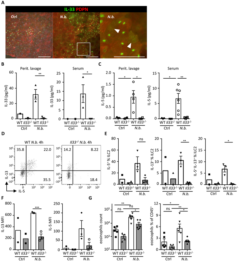
Obesity is driven by chronic low-grade inflammation resulting from dysregulated immune cell accumulation and function in white adipose tissue (WAT). Interleukin-33 (IL-33) is a key cytokine that controls innate and adaptive immune cell activity and immune homeostasis in WAT, although the sources of IL-33 have remained controversial. Here, we show that WAT-resident mesenchyme-derived stromal cells are the dominant producers of IL-33. Adipose stem and progenitor cells (ASPCs) produced IL-33 in all WAT depots, whereas mesothelial cells served as an additional source of IL-33 in visceral WAT. ASPC-derived IL-33 promoted a regulatory circuit that maintained an immune tone in WAT via the induction of group 2 innate lymphoid cell-derived type 2 cytokines and maintenance of eosinophils, whereas mesothelial IL-33 also acted as an alarmin by inducing peritoneal immune response upon infection. Together, these data reveal a previously unrecognized regulatory network between tissue-resident progenitor cells and innate lymphoid cells that maintains immune homeostasis in adipose tissue.


Figures

Fig. 1.
Fig. 1.
IL-33 regulates ILC2 activity and eosinophil numbers. (A) Body weight of wild type (WT) and Il33−/− mice on conventional diet at 16 weeks of age. (n = 4) (B) Mass of indicated adipose depots in WT and Il33−/− mice at 16 weeks of age. (n = 4) (C) Proportion of combined white adipose tissue (WAT) mass of epididymal (ewat), mesenteric (mwat) and inguinal (iwat) depots as percent of body weight in WT and Il33−/− mice at 16 weeks of age. (n = 4) (D) ILC2 numbers and proportions in visceral adipose compartments of WT and Il33−/− mice. (n ≥ 3) Data pooled from two individual experiments. (E and F) Representative dot plots (E) and quantification (F) showing intracellular IL-5 and IL-13 staining in ILC2s from visceral adipose depots of WT and Il33−/− mice. (n = 3) (G and H) Representative dot plots (G) and quantification (H) of macrophages and eosinophils in visceral adipose depots of WT and Il33−/− mice. (n = 3). Mean ± SEM, * P < 0.05, ** P < 0.01, ***P < 0.001, ns - not significant, Student’s t-test.
Fig. 2.
Fig. 2.
Visceral WAT adipose stem and progenitor cells produce IL-33. (A and B) Expression of Il33 in (A) isolated adipocytes (adip.), stromal vascular fraction (SVF) and in (B) indicated sort-purified cells of murine epididymal WAT (ewat) as determined by qRT-PCR analysis. (n ≥ 5) (C) Representative histogram overlay and dot plots of cells in ewat SVF fraction of wild type mice, stained with anti-IL-33 (α-IL-33) and IgG control (ctrl) antibodies. Data depicted are gated on live cells. Numbers in gates indicate proportion (%) of parent gate. (D) Histogram demonstrating mean normalized counts from RNAseq of genes expressed in sort-purified ASPCs from ewat. Markers of ASPCs are indicated in black, highly expressed cytokine genes are indicated in blue, and Il33 is shown in red. Genes with mean normalized counts < 10 are not shown. (n = 3) (E) Expression of Il33 in adipocytes and sort-purified cells from ewat as determined by qRT-PCR analysis. (n ≥ 5) (F) Representative dot plots showing eGFP-expressing cells in ewat of the IL-33-eGFP mouse. Numbers in gates indicate proportion (%) of parent gate. Data shown are gated on live cells. (G) Representative dot plot and histogram overlay of live eGFP+ lin (CD31+ CD45+ Ter119+) cells in the ewat of the IL-33-eGFP mouse. (H) Expression of indicated genes in isolated adipocytes and sort-purified cells from the ewat as determined by qRT-PCR analysis. (n ≥ 5) (I) Representative dot plots of mesothelin (MSLN) staining in wild type mouse SVF from ewat and subcutaneous WAT (inguinal WAT; iwat) first gated on live lin cells. Numbers in gates indicate proportion (%) of the parent gate. (J) Quantification of eGFP+ cell populations in indicated WAT compartments. Data were compiled from three independent experiments (n ≥ 13). Mean ± SEM, * P < 0.05, ** P < 0.01, ***P < 0.001, ****P < 0.0001, ns - not significant, Student’s t-test.
Fig. 3.
Fig. 3.
Localization of IL-33-expressing cells in visceral WAT. (A) Surface view of the ewat from the IL-33-eGFP reporter mouse (right) and a wild type (WT) control animal (left). (B) Immunostaining of IL-33 (green) in paraffin-embedded WT mouse ewat. Note IL-33 localization to DAPI-stained nuclei (blue) scattered between perilipin+ adipocytes (purple), as well as IL-33 in the PDPN+ (red) membrane lining the adipose compartment. (C) Immunostaining of paraffin-embedded WT mouse ewat. PDGFRα-expressing ASPCs (green) embedded between perilipin+ adipocytes (purple). (D) Immunostained transverse sections (side view) of whole-mount WT mouse ewat demonstrating PDGFRα-stained (purple) ASPCs that express IL-33 (green) in the nucleus (blue). The PDPN-stained (red) cell layer expressing nuclear IL-33 (green) envelops the adipose compartment. Arrowheads indicate magnified areas in adjacent panels. (E) Side and surface view of the same sample of WT mouse whole-mount stained ewat. (F and G) Surface view of whole-mount stained ewat. ZO1 (green) staining on PDPN+ (red) cobblestone-shaped cells in (F) and co-localization of IL-33 (green) and WT1 (purple) in the nucleus (blue) in (G). Scale bar, 100 μm, unless indicated otherwise. All immunostaining experiments are representative of two mice, and the staining protocol has been repeated minimum of three times.
Fig. 4.
Fig. 4.
Adipose stem and progenitor cell-derived IL-33 controls ILC2 activity. (A) Representative histogram and dot plots showing ST2+ cells in the lymphocyte population first gated on live CD45+ cells. (B) Representative histogram of ST2 expression on ILC2 (defined as shown in fig. S9A) from the small intestinal lamina propria (SI LP) and epididymal WAT (ewat). (B and C) Representative histogram (B) and bar graph (C) showing mean fluorescence intensity (MFI) of ST2 on ILC2s from ewat and SI LP. (n = 6) (D) Schematic indicating experimental design for E and F. (E) Sort-purified ILC2s from wild type (WT) or Il1rl1−/− mouse visceral WAT (pooled ewat and mwat) ILC2s were cultured in ASPC-conditioned (ASPC) or control media (Ctrl) and IL-5 secretion was analyzed by ELISA after 48 h. (n = 3) (F) Sort-purified ILC2s from visceral WAT or SI LP were cultured in ASPC-conditioned or control media (Ctrl) and IL-5 and IL-13 secretion was analyzed by multiplex bead-based assay after 48 h. (n = 3) (G) Schematic indicating experimental design. Sort-purified ASPCs from wild type (WT) or Il33−/− mice were injected into the peritoneum of Il33−/− mice and immunological analysis (H-J) performed after six weeks in visceral adipose compartments. (H) ILC2 numbers, (I) proportions of IL-5+ and IL-13+ ILC2s, and (J) eosinophil quantification. (n = 4). Mean ± SEM, * P < 0.05, ** P < 0.01, ***P < 0.001, ns – not significant, Student’s t-test.
Fig. 5.
Fig. 5.
IL-33 controls adipose tissue expansion and immunological homeostasis in short-term HFD feeding. (A-B) Wild type mice were fed with high-fat diet (HFD) or control diet (CD) up to 7 days (d). (A) Representative dot plot of Ki67 staining in ASPCs (defined as live lin (CD31 CD45 Ter119)) PDGFRα+Sca-1+ cells) on day 7. (B) Proportion of proliferating cells (Ki67+) cells among total ASPCs from indicated WAT depots. (n = 3) (C) Expression of Il33 and Mcp1 in sort-purified ASPCs isolated from ewat of wild type mice (WT) on CD or HFD for 3 days as determined by qRT-PCR. (n = 3) (D to I) WT or Il33−/− mice were fed with CD or HFD and sacrificed for analysis after 3 days. (D) Body weight change as a proportion (%) of initial weight (day 0). Data compiled from two individual experiments. (n ≥ 7) (E) Proportion of ewat mass as proportion (%) of body weight. Data compiled from two individual experiments. (n ≥ 7) (F) Proportion of Ki67+ and adipocyte progenitor cells (CD24+) among total ewat ASPCs. (G) Mean fluorescence intensity (MFI) of forward scatter (FSC) and side scatter (SSC) of ewat ASPCs. (H and I) Quantification of macrophages (H) and dendritic cells (DCs) (I) in ewat. (n ≥ 4). Mean ± SEM, * P < 0.05, ** P < 0.01, ***P < 0.001, Student’s t-test.
Fig. 6.
Fig. 6.
Mesothelial IL-33 acts as an alarmin to activate the immune system upon peritoneal infection. (A) Wild type (WT) mice were injected i.p. with Nippostrongylus brasiliensis (N.b.) L3 larvae and epididymal WAT (ewat) extracted after 1 h. Representative surface view of immunostained whole-mount ewat depicting IL-33 (green) and PDPN+ mesothelium (red). Note IL-33 relocalization from the nucleus to the cytoplasm in N.b.-treated condition. Encircled area indicates magnified area. Arrowheads point to diffuse IL-33 staining in the cytoplasm. (B and C) ELISA quantification of IL-33 and IL-5 in peritoneal lavage fluid and serum of wild type (WT) and Il33−/− mice 4 h after N.b. infection. (n ≥ 3) (D - E) Representative dot plots (D) and quantification (E) showing intracellular IL-5 and IL-13 staining in ILC2s from ewat of WT and Il33−/− mice treated with N.b. (n ≥ 2) (F) Mean fluorescence intensity (MFI) of IL-13 and IL-5 in ILC2s from panels D and E. (G) Quantification of eosinophils in peritoneal lavage fluid of WT and Il33−/− mice treated with N.b. at 4 h. (n ≥ 5) Mean ± SEM, * P < 0.05, ** P < 0.01, ***P < 0.001, ns - not significant, Student’s t-test.

Comment in

  • Stromal IL-33 balances fat stores.
    Bird L. Bird L. Nat Rev Immunol. 2019 Jul;19(7):412-413. doi: 10.1038/s41577-019-0179-7. Nat Rev Immunol. 2019. PMID: 31110269 No abstract available.

References

    1. Hales CM, Carroll MD, Fryar CD, Ogden CL, Prevalence of Obesity Among Adults and Youth: United States, 2015–2016. NCHS Data Brief, 1–8 (2017). - PubMed
    1. Gonzalez-Muniesa P et al., Obesity. Nat Rev Dis Primers 3, 17034 (2017). - PubMed
    1. Brestoff JR, Artis D, Immune regulation of metabolic homeostasis in health and disease. Cell 161, 146–160 (2015). - PMC - PubMed
    1. Melen E et al., Analyses of shared genetic factors between asthma and obesity in children. J Allergy Clin Immunol 126, 631–637 e631–638 (2010). - PMC - PubMed
    1. Angeles-Martinez J et al., The rs7044343 Polymorphism of the Interleukin 33 Gene Is Associated with Decreased Risk of Developing Premature Coronary Artery Disease and Central Obesity, and Could Be Involved in Regulating the Production of IL-33. PLoS One 12, e0168828 (2017). - PMC - PubMed

Publication types

MeSH terms