. 2019 Jul:89:99-108.
doi: 10.1016/j.humpath.2019.04.009. Epub 2019 May 3.

Tissue-specific quantification and localization of androgen and estrogen receptors in prostate cancer

Affiliations

Tissue-specific quantification and localization of androgen and estrogen receptors in prostate cancer

Priyanka D Sehgal et al. Hum Pathol. 2019 Jul.

Abstract

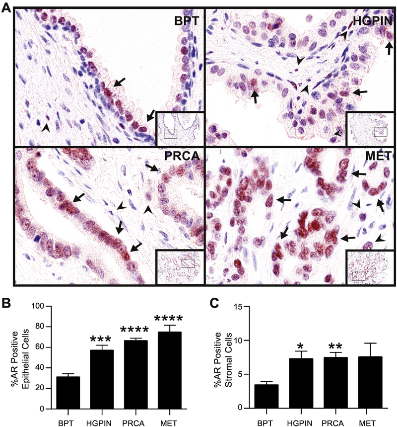
Androgens and estrogens, working together, promote prostate cancer (PRCA) initiation and progression, with androgens acting via androgen receptor (AR) and estrogens acting primarily through estrogen receptor α (ERα). While the interplay between these steroid hormones has been established, the interaction between steroid hormone receptors in prostatic disease remains unstudied. The goal of this study was to objectively determine the incidence, stage specificity, and tissue/cell type specificity of AR and ERα expression, both independently and simultaneously, during the progression of PRCA. Using multiplexed immunohistochemistry and multispectral imaging analysis, AR, ERα, and smooth muscle α-actin expression was detected and quantitated in benign prostate tissue (BPT), high-grade prostatic intraepithelial neoplasia (HGPIN), PRCA, and metastasis (MET) from patient specimens (n=340). Epithelial AR expression was significantly increased in HGPIN, PRCA, and MET compared with BPT, whereas ERα expression in epithelial and stromal cells was highest in HGPIN. With analysis of AR and ERα coexpression, we identified a unique population of double-positive (AR+/ERα+) cells that increased in HGPIN specimens in both the stroma and the epithelium. Double-negative (AR-/ERα-) cells significantly decreased across PRCA progression, from 65% in BPT to 30% in MET. Preliminary analysis of this AR+/ERα+ population indicates potential cell type specificity in smooth muscle α-actin-negative stromal cells. This study demonstrates stage-, tissue-, and cell type-specific AR and ERα expression changes during PRCA progression, both independently and coexpressed. A more complete understanding of steroid hormones and their receptors in the initiation and progression of prostatic disease may elucidate improved strategies for PRCA prevention or therapy.

Keywords: Androgens; Epithelium; Estrogens; Prostate cancer; Stroma.


Figures

Fig. 1.
Fig. 1.. Androgen receptor expression in prostate cancer progression.
A: Prostate composite image shows androgen receptor (AR, red) in nuclei of luminal epithelial cells (arrows) and stromal cells (arrowheads). AR positivity is seen more in the epithelium than stroma, and overall, expression increased in PRCA progression. B: The percentage of AR positive cells significantly increased in the epithelium in HGPIN, PRCA and MET compared to BPT. C: The percentage of AR positive cells in the stroma significantly increased in HGPIN and PRCA compared to BPT. BPT = benign tissue prostate, HGPIN = high grade prostatic neoplasia, PRCA = prostate cancer, MET = metastasis. IHC 100X (20X inset). *P < 0.05, **P < 0.01, ***P < 0.001, ****P < 0.0001 via post-hoc comparison to BPT.
Fig. 2.
Fig. 2.. Estrogen receptor-alpha expression in prostate cancer progression.
A: Prostate composite image shows estrogen receptor-alpha (ERα, brown) in nuclei of epithelial (arrows) and stromal cells (arrow heads). In normal prostate epithelium, ERα was localized primarily in basal epithelial cells, while epithelial ERα expression in HGPIN and PRCA showed a marked increase. B: The overall percentage of ERα positive cells in the epithelium is significantly increased in HGPIN compared to BPT. C: The percentage of ERα positive stromal cells was increased in HGPIN and PRCA compared to BPT. BPT = benign tissue prostate, HGPIN = high grade prostatic neoplasia, PRCA = prostate cancer, MET = metastasis. IHC 100X (20X inset). *P < 0.05, **P < 0.01, ****P < 0.0001 via post-hoc comparison to BPT.
Fig. 3.
Fig. 3.. Co-localization of AR and ERα in normal and pathologic prostate.
A: Prostate composite image shows a small number of AR+/ERα+ double positive nuclei (yellow) seen within the epithelium (arrow) and stroma (arrowheads) in all stages of PRCA progression. B: The percent positivity of AR+/ERα+ cells in the epithelium was increased in HGPIN only compared to BPT. C: Double negative (AR/ERα) cells in the epithelium were significantly decreased in HGPIN, PRCA and MET compared to BPT. D: In the stroma, AR+/ERα+ double positive cells were significantly higher in HGPIN compared to BPT. E: Double negative (AR/ERα) cells in the stroma were significantly decreased in HGPIN and PRCA compared to BPT, but unchanged in MET compared to BPT. BPT = benign tissue prostate, HGPIN = high grade prostatic neoplasia, PRCA = prostate cancer, MET = metastasis. IHC 100X (20X inset). **P < 0.01, ***P < 0.001, ****P < 0.0001 via post-hoc comparison to BPT.
Fig. 4.
Fig. 4.. AR and ERα expression and co-localization in smooth muscle α-actin negative stroma.
A: Prostate composite image shows localization of AR (red) and smooth muscle α-actin (SMA, turquoise) in PRCA progression. Quantification of AR in SMA-negative stroma showed a significant increase in HGPIN and PRCA compared to BPT, but a significant decrease in MET compared to BPT. B: Prostate composite image shows localization of ERα (brown) and smooth muscle alpha-actin (SMA, turquoise) in PRCA progression. Quantification of AR in SMA-negative stroma showed a significant increase in HGPIN compared to BPT, and no significant change in PRCA or MET compared to BPT. C: Prostate composite image shows co-localization of cells expressing both AR and ERα (yellow) and smooth muscle α-actin (SMA, turquoise) in PRCA progression. Quantification of AR+/ERα+ cells in SMA-negative stroma showed a significant increase in HGPIN compared to BPT, and no significant change in PRCA or MET compared to BPT. BPT = benign tissue prostate, HGPIN = high grade prostatic neoplasia, PRCA = prostate cancer, MET = metastasis. IHC 100X (20X inset). *P < 0.05, **P < 0.01 via post-hoc comparison to BPT.

References

    1. Siegel RL, Miller KD, Jemal A. Cancer statistics, 2019. CA: a cancer journal for clinicians. - PubMed
    1. Prins GS, Korach KS. The role of estrogens and estrogen receptors in normal prostate growth and disease. Steroids 2008; 73:233–44. - PMC - PubMed
    1. Ricke WA, Wang Y, Cunha GR. Steroid hormones and carcinogenesis of the prostate. the role of estrogens. Differentiation 2007; 75:871–82. - PubMed
    1. Ricke WA, McPherson SJ, Bianco JJ, Cunha GR, Wang Y, Risbridger GP. Prostatic hormonal carcinogenesis is mediated by in situ estrogen production and estrogen receptor alpha signaling. The FASEB journal 2008; 22:1512–20. - PubMed
    1. Fujimura T, Takayama K, Takahashi S, Inoue S. Estrogen and androgen blockade for advanced prostate cancer in the era of precision medicine. Cancers 2018; 10:29. - PMC - PubMed

Publication types