. 2019 Jun 20;74(6):1164-1174.e4.
doi: 10.1016/j.molcel.2019.04.008. Epub 2019 May 1.

Crosstalk between RNA Pol II C-Terminal Domain Acetylation and Phosphorylation via RPRD Proteins

Affiliations

Crosstalk between RNA Pol II C-Terminal Domain Acetylation and Phosphorylation via RPRD Proteins

Ibraheem Ali et al. Mol Cell. .

Abstract

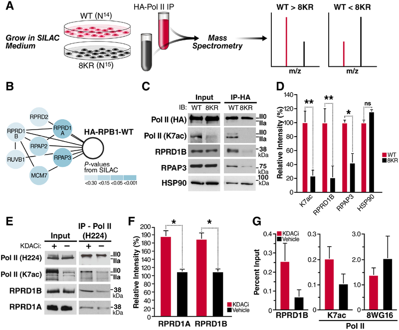
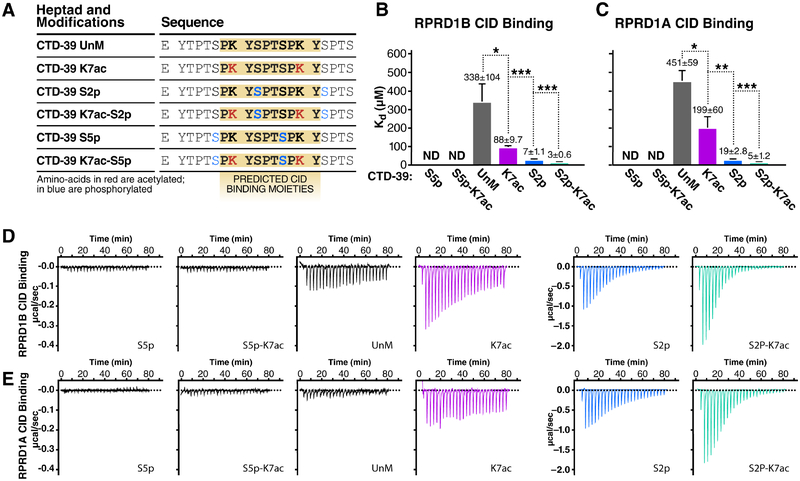
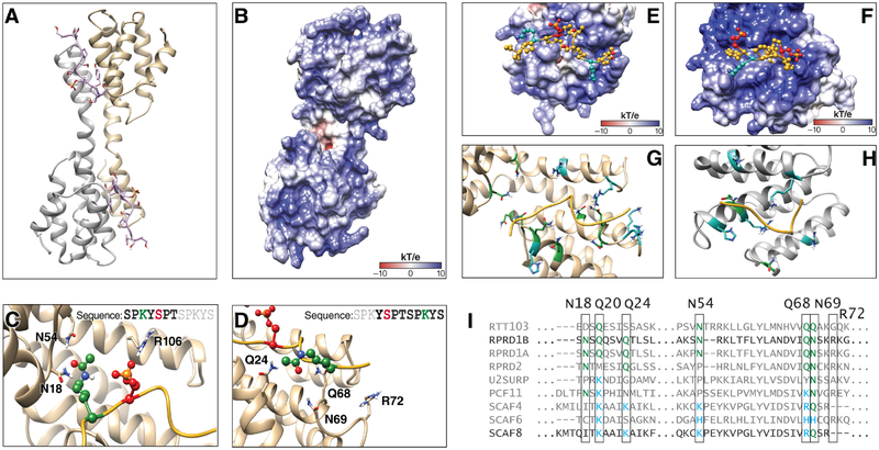
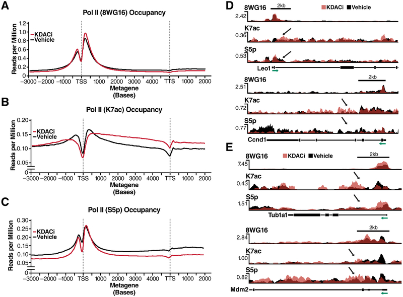
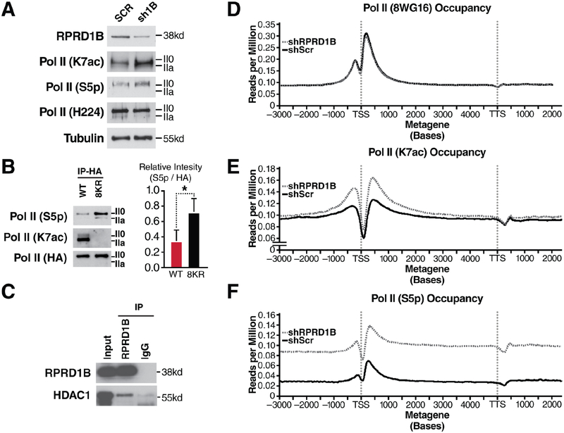
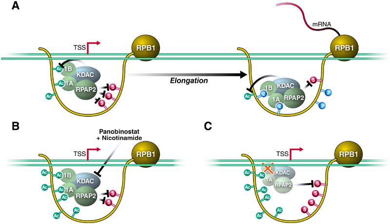
Post-translational modifications of the RNA polymerase II C-terminal domain (CTD) coordinate the transcription cycle. Crosstalk between different modifications is poorly understood. Here, we show how acetylation of lysine residues at position 7 of characteristic heptad repeats (K7ac)-only found in higher eukaryotes-regulates phosphorylation of serines at position 5 (S5p), a conserved mark of polymerases initiating transcription. We identified the regulator of pre-mRNA-domain-containing (RPRD) proteins as reader proteins of K7ac. K7ac enhanced CTD peptide binding to the CTD-interacting domain (CID) of RPRD1A and RPRD1B proteins in isothermal calorimetry and molecular modeling experiments. Deacetylase inhibitors increased K7ac- and decreased S5-phosphorylated polymerases, consistent with acetylation-dependent S5 dephosphorylation by an RPRD-associated S5 phosphatase. Consistent with this model, RPRD1B knockdown increased S5p but enhanced K7ac, indicating that RPRD proteins recruit K7 deacetylases, including HDAC1. We also report autoregulatory crosstalk between K7ac and S5p via RPRD proteins and their interactions with acetyl- and phospho-eraser proteins.

Keywords: C-terminal domain; Pol II CTD; RNA polymerase II; acetylation; crosstalk; gene regulation; histone deacetylase; phosphorylation; post-translational modification; transcription.


Conflict of interest statement

DECLARATION OF INTERESTS

The authors declare no competing interests.

Figures

Figure 1:
Figure 1:. RPRD Proteins Interact with RPB1 in an Acetylation-Dependent Manner.
(A) A SILAC-based mass spectrometry screen was used to identify factors that bind preferentially to WT Pol II, compared to the 8KR mutant. (B) Components of the RPRD1B/RPRD1A complex that were identified as WT interactors. (C) Hemagglutinin (HA) immunoprecipitation and western blotting with the indicated antibodies RPRD1B or from WT and 8KR cells. (D) Densitometry using ImageJ of at least three independent western blots from indicated antibodies after HA-immunoprecipitation. (E) NIH3T3 cells were treated with KDAC inhibitors (30 nM Panobinostat and 5 μM Nicotinamide) for 2 h. IP using an N-terminal H224 antibody from 500 μg of nucleoplasm pre-cleared with IgG and western blotting with the indicated antibodies. (F) Densitometry of RPRD1A and RPRD1B western blots produced from three independent Pol II Total (H224) IP elutions. (G) ChIP-qPCR on the Leo1 gene at +33 nt downstream of TSS using the indicated antibodies from 4 independent chromatin preparations. Unmodified Pol II ChIP experiments were done with the 8WG16 antibody. Values are represented as percent of input with IgG subtracted. Data is shown as mean ± SEM. *p < 0.5; **p < 0.01; ns, not significant for a one-tailed T test. See also Table S1.
Figure 2:
Figure 2:. RPRD CID Domains Recognize Acetylated and Phosphorylated CTD Peptides.
Isothermal titration calorimetry (ITC) experiments measuring in-vitro binding affinity between RPRD1A and RPRD1B CID domains and modified CTD peptides. (A) Table of CTD-39 peptides with indicated post-translational modifications and predicted RPRD CID binding moieties. (B) Kd values measured by ITC for RPRD1B CID and modified CTD-39 binding. (C) Kd values measured by ITC for RPRD1A CID and modified CTD-39 binding. (D) Representative ITC plots showing effect of K7ac on RPRD1B CID-CTD interactions for the indicated modifications (E) Representative ITC plots showing effect of K7ac on RPRD1A CID-CTD interactions for the indicated modifications. Data is shown as mean ± SEM. * p < 0.05; ** p < 0.01; *** p < 0.005 using a one-tailed T test.
Figure 3:
Figure 3:. Molecular Modeling of K7 Acetylation and Interaction with CID Domains.
(A) Dimer model from RPRD1B crystal structure (pdb:4Q94) containing two recognition modules and two peptides fragments of the CTD. (B) Electrostatic potential surface for RPRD1B CID. (C) Recognition elements around first K7ac and S2p in the CTD peptide fragment. (D) Recognition elements around second K7ac in the CTD peptide fragment. (E) CTD peptide fragment model from crystal structure superimposed with the electrostatic surface potential around the corresponding binding site. (F) CTD peptide fragment superimposed with the electrostatic potential surface around the corresponding SCAF8 CID binding site (G) RPRD1B CID residues found within 5Ǻ of peptide fragment. Blue: positively charged residues. Green: amide-containing residues. (H) SCAF8 CID residues found within 5Ǻ of peptide fragment. Blue: positively charged residues. Green: amide-containing residues. (I) Clustal Omega sequence alignment of human CID regions with yeast RTT103 as outgroup. Green: amide-containing residues. Blue: positively charged residues. See also Figure S1.
Figure 4:
Figure 4:. An Inverse Relationship between K7ac and Phosphorylation Is Induced upon KDAC Inhibition.
(A-C) Metagene profiles generated from chromatin immunoprecipitation experiments in NIH3T3 cells with the indicated antibodies followed by deep-sequencing. Gene profiles are measured in reads per million normalized to input control (D) Single-gene validation of RNA Pol II PTMs measured as reads per million on selected RPRD1B occupied genes. S5p was measured using the RNA Pol II 4H8 antibody. (E) Occupancy profiles of RNA Pol II PTMs on control genes. Green arrows indicate the direction of transcription relative to the TSS of the depicted gene. Black arrows indicate the site of affected PTMs in response to KDACi.
Figure 5:
Figure 5:. Increased S5p and K7ac Levels in Response to RPRD1B Knockdown.
(A) Western blotting of indicated RNA Pol II modifications in NIH3T3 cells treated with scrambled shRNAs or those targeting RPRD1B. (B) Western blotting against S5p and K7ac from HA-immunopurified lysates containing WT or 8KR RNA Pol II and densitometry using ImageJ of three independent HA-elutions. (C) RPRD1B immunoprecipitation from 293T cell lysates and western blotting against nuclear histone deacetylase HDAC1. (D-F) Metagene profiles of expressed genes in NIH3T3 cells. Gene profiles are measured in reads per million, relative to input control. Metagene profiles were generated from ChIP-seq data from NIH3T3 cells expressing either scrambled or RPRD1B targeted shRNAs treated with vehicle control. S5p is measured using the RNA Pol II 4H8 antibody. Profiles are representative of two independent experiments. Data is shown as mean ± SEM. * p < 0.05; using a one-tailed T test. See also Figure S2 and Table S2.
Figure 6:
Figure 6:. RPRD Proteins Are Recruited to the RPB1 CTD via Acetylation and Phosphorylation to Antagonize S5-Phosphorylation.
(A) Model of RPRD complex reader and effector functions along modified residues within the CTD. 1B- RPRD1B 1A- RPRD1A, KDAC represents RPRD associated K7 deacetylases. (B) KDAC inhibition induces K7 hyperacetylation and downregulation of S5-phosphorylation. (C) Knockdown of RPRD1B perturbs the recruitment of the complex to result in both S5 hyperphosphorylation and K7 hyperacetylation. See text for details.

References

    1. BARTKOWIAK B, LIU P, PHATNANI HP, FUDA NJ, COOPER JJ, PRICE DH, ADELMAN K, LIS JT & GREENLEAF AL 2010. CDK12 is a transcription elongation-associated CTD kinase, the metazoan ortholog of yeast Ctk1. Genes Dev, 24, 2303–16. - PMC - PubMed
    1. BARTOLOMEI MS & CORDEN JL 1987. Localization of an alpha-amanitin resistance mutation in the gene encoding the largest subunit of mouse RNA polymerase II. Mol Cell Biol, 7, 586–94. - PMC - PubMed
    1. BASTA J & RAUCHMAN M 2015. The nucleosome remodeling and deacetylase complex in development and disease. Transl Res, 165, 36–47. - PMC - PubMed
    1. BECKER R, LOLL B & MEINHART A 2008. Snapshots of the RNA processing factor SCAF8 bound to different phosphorylated forms of the carboxyl-terminal domain of RNA polymerase II. J Biol Chem, 283, 22659–69. - PubMed
    1. BROOKES E, DE SANTIAGO I, HEBENSTREIT D, MORRIS KJ, CARROLL T, XIE SQ, STOCK JK, HEIDEMANN M, EICK D, NOZAKI N, KIMURA H, RAGOUSSIS J, TEICHMANN SA & POMBO A 2012. Polycomb associates genome-wide with a specific RNA polymerase II variant, and regulates metabolic genes in ESCs. Cell Stem Cell, 10, 157–70. - PMC - PubMed

Publication types

MeSH terms