. 2019 Jun 15:409:195-203.
doi: 10.1016/j.neuroscience.2019.04.045. Epub 2019 May 2.

A sexually dimorphic distribution of corticotropin-releasing factor receptor 1 in the paraventricular hypothalamus

Affiliations

A sexually dimorphic distribution of corticotropin-releasing factor receptor 1 in the paraventricular hypothalamus

Zachary J Rosinger et al. Neuroscience. .

Erratum in

Abstract

Sex differences in neural structures are generally believed to underlie sex differences reported in anxiety, depression, and the hypothalamic-pituitary-adrenal axis, although the specific circuitry involved is largely unclear. Using a corticotropin-releasing factor receptor 1 (CRFR1) reporter mouse line, we report a sexually dimorphic distribution of CRFR1 expressing cells within the paraventricular hypothalamus (PVN; males > females). Relative to adult levels, PVN CRFR1-expressing cells are sparse and not sexually dimorphic at postnatal days 0, 4, or 21. This suggests that PVN cells might recruit CRFR1 during puberty or early adulthood in a sex-specific manner. The adult sex difference in PVN CRFR1 persists in old mice (20-24 months). Adult gonadectomy (6 weeks) resulted in a significant decrease in CRFR1-immunoreactive cells in the male but not female PVN. CRFR1 cells show moderate co-expression with estrogen receptor alpha (ERα) and high co-expression with androgen receptor, indicating potential mechanisms through which circulating gonadal hormones might regulate CRFR1 expression and function. Finally, we demonstrate that a psychological stressor, restraint stress, induces a sexually dimorphic pattern of neural activation in PVN CRFR1 cells (males >females) as assessed by co-localization with the transcription/neural activation marker phosphorylated CREB. Given the known role of CRFR1 in regulating stress-associated behaviors and hormonal responses, this CRFR1 PVN sex difference might contribute to sex differences in these functions.

Keywords: androgen; corticotropin releasing factor; sex difference; stress.


Figures

Figure 1.
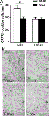
Figure 1.. Sex difference in CRFR1-GFP cells in the PVN.
(A) Male mice have a greater number of CRFR1-GFP containing cells in the PVN compared to females. (B) Representative images of CRFR1-GFP cells in an adult (P60) male and female mouse brain. *p≤ 0.01. 3v; 3rd ventricle.
Figure 2.
Figure 2.. CRFR1-GFP cell number in the P0, P4, and P21 mouse PVN.
(A) No sex differences were found in CRFR1-GFP within the PVN at P0, P4, or P21. Relative to adult levels, CRFR1-GFP was low at these postnatal time points. (B) Representative images showing CRFR1-GFP in the P0, P4, and P21 PVN. Dashed lines approximate anatomical borders of the PVN (Paxinos et al., 2007; Allen Institute Mouse Reference Atlas) 3v; 3rd ventricle.
Figure 3.
Figure 3.. CRFR1-GFP cell number in the male and female 20–24 month old mouse.
In 20–24 month old mice, the adult sex difference in CRFR1-GFP (males > females) persists. *p≤ 0.05.
Figure 4.
Figure 4.. Gonadectomy effects on PVN CRFR1-GFP cell number.
(A) Orchidectomy in adult males results in a decrease in CRFR1-GFP cell number while ovariectomy in females produces no effects on CRFR1-GFP cell number. (B) Representative images of PVN CRFR1-GFP cells in sham and gonadectomized males and females. * indicates p≤0.01. 3v; 3rd ventricle. GDX; gonadectomy.
Figure 5.
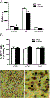
Figure 5.. Co-localization of CRFR1-GFP with estrogen receptor alpha (ERα) in the PVN.
(A) A male/female comparison of CRFR1-GFP, ERα, co-localized CRFR1-GFP/ERα, and (B) the percentage of CRFR1-GFP/ERα co-localized cells in the PVN. Approximately 29% of CRFR1 cells were found to be co-localized with ERα although no sex differences in the number or percentage of co-localized cells were found. No differences were found for the percentage of CRFR1 cells co-expressing ERα between the rostral (r) and middle (m) PVN. (C-H) High and low magnification representative images of a male mPVN showing co-localization of CRFR1-GFP and ERα. Note the distinct distribution overlap for CRFR1-GFP and ERα in the low magnification images (F-H). Arrows indicate examples of co-localized cells. * indicates p≤0.05. 3v; 3rd ventricle.
Figure 6.
Figure 6.. Co-localization of CRFR1-GFP with androgen receptor (AR) in the PVN.
(A) CRFR1-GFP, AR, co-localized CRFR1-GFP/AR, and (B) the percentage of CRFR1-GFP/AR colocalized cells in the male and female PVN. Approximately 65% of CRFR1 cells co-localized with AR with no sex differences in the number or percentage of co-localized cells found. No differences were found for the percentage of CRFR1 cells co-expressing AR between the rostral (r) and middle (m) PVN (B). A low magnification (C) and high magnification image of middle PVN showing co-localized cells containing black AR label within brown CRFR1-GFP cells (D). Arrows indicate co-localized cells. # indicates p=0.08 compared to males. * indicates p≤0.05 compared to males. 3v; 3rd ventricle.
Figure 7.
Figure 7.. Co-localization of CRFR1-GFP with phosphorylated Creb (pCREB) following restraint stress.
(A) Males show a greater number of CRFR1-GFP cells that co-express pCREB following a 30 minute restraint stress. (B) The percentage of cells that co-express CRFRI-GFP/pCREB did not differ by sex. (C-D) Representative images of a male PVN showing co-localization of CRFR1-GFP and pCREB in a stressed and unstressed mouse. Arrows indicate examples of co-localized cells. * indicates p≤0.01. 3v; 3rd ventricle.

References

    1. Abel KB, Majzoub JA (2005), Molecular biology of the HPA axis. Ed. Steckler T, Kalin NH, and Reul JMHM. Techniques in Behavioral and Neural Sciences 15(1):79–94.
    1. Bale TL, Picetti R, Contarino A, Koob GF, Vale WW, Lee KF (2002), Mice deficient for both corticotropin-releasing factor receptor 1 (CRFR1) and CRFR2 have an impaired stress response and display sexually dichotomous anxiety-like behavior. J Neurosci 22(1):193–199. - PMC - PubMed
    1. Bangasser DA (2013), Sex differences in stress-related receptors: “micro” differences with “macro” implications for mood and anxiety disorders. Biol Sex Differ 4(1):2. - PMC - PubMed
    1. Bangasser DA, Curtis A, Reyes BA, Bethea TT, Parastatidis I, Ischiropoulos H, Van Bockstaele EJ, Valentino RJ (2010), Sex differences in corticotropin-releasing factor receptor signaling and trafficking: potential role in female vulnerability to stress-related psychopathology. Mol Psychiatry 15(9):877, 896–904. - PMC - PubMed
    1. Bangasser DA, Eck SR, Ordones Sanchez E (2019), Sex differences in stress reactivity in arousal and attention systems. Neuropsychopharmacology 44(1):129–139. - PMC - PubMed

Publication types

MeSH terms

Substances