. 2019 Sep 1;1865(9):2210-2223.
doi: 10.1016/j.bbadis.2019.04.018. Epub 2019 May 2.

COL1A1 C-propeptide mutations cause ER mislocalization of procollagen and impair C-terminal procollagen processing

Affiliations

COL1A1 C-propeptide mutations cause ER mislocalization of procollagen and impair C-terminal procollagen processing

Aileen M Barnes et al. Biochim Biophys Acta Mol Basis Dis. .

Abstract

Mutations in the type I procollagen C-propeptide occur in ~6.5% of Osteogenesis Imperfecta (OI) patients. They are of special interest because this region of procollagen is involved in α chain selection and folding, but is processed prior to fibril assembly and is absent in mature collagen fibrils in tissue. We investigated the consequences of seven COL1A1 C-propeptide mutations for collagen biochemistry in comparison to three probands with classical glycine substitutions in the collagen helix near the C-propeptide and a normal control. Procollagens with C-propeptide defects showed the expected delayed chain incorporation, slow folding and overmodification. Immunofluorescence microscopy indicated that procollagen with C-propeptide defects was mislocalized to the ER lumen, in contrast to the ER membrane localization of normal procollagen and procollagen with helical substitutions. Notably, pericellular processing of procollagen with C-propeptide mutations was defective, with accumulation of pC-collagen and/or reduced production of mature collagen. In vitro cleavage assays with BMP-1 ± PCPE-1 confirmed impaired C-propeptide processing of procollagens containing mutant proα1(I) chains. Overmodified collagens were incorporated into the matrix in culture. Dermal fibrils showed alterations in average diameter and diameter variability and bone fibrils were disorganized. Altered ER-localization and reduced pericellular processing of defective C-propeptides are expected to contribute to abnormal osteoblast differentiation and matrix function, respectively.

Keywords: BMP-1; C-propeptide; Collagen processing; Endoplasmic reticulum localization; Osteogenesis imperfecta.


Conflict of interest statement

Disclosure statement: The authors declare no conflicts of interest.

Figures

Fig. 1.
Fig. 1.
Structural model of the CPI heterotrimer and positions of mutation sites. A) Overall structure showing the stalk, base and petal regions, the two α1(I) chains in beige, the α2(I) chain in green, bound Ca2+ ions in blue and disulfide bonds in yellow. Mutation sites are indicated (in bold) for one of the two α1(I) chains. Residues are numbered from the N-terminus of the C-propeptide after cleavage from the rest of the molecule, as well as from the transcription start site of the full-length chain (in parentheses). B) Close-up of W57(1275), G63(1281) and other residues in the base region. C) Close-up of T80(1298) and neighboring disulfide bonds at the base/petal junction. D) Close-up of D195(1413), D223(1441) and P226(1444) and a nearby disulfide bond in the petal region. Residues marked with an apostrophe (‘) refer to adjacent chains in the trimeric structure.
Fig. 2.
Fig. 2.
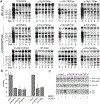
Probands with C-propeptide mutations have overmodified steady-state collagen protein. A) Autoradiography of type I collagen chains from all probands shows backstreaking or overmodification of the α1(I) and α2(I) chains on SDS-urea-PAGE; slower migrating forms were more prominent in the cell fraction. B) Differential scanning calorimetry of C-propeptide probands’ collagens shows normal thermal stability.
Fig. 3.
Fig. 3.
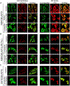
Procollagen with C-propeptide mutations dissociates from the rER membrane and localizes to the ER lumen. A) Immunofluorescence microscopy shows colocalization of the C-propeptide and the ER membrane-bound chaperone calnexin in control fibroblasts and fibroblasts from a patient with the helical mutation p.Gly1175Ser. C-propeptide (p.Thr1298Ile) proband fibroblasts show decreased overlap of the C-propeptide with calnexin and an increased overlap with the ER luminal chaperone PDI. B) Immunofluorescence microscopy of fibroblasts with mutations in the petal region of the C-propeptide shows large C-propeptide aggregates (white arrowheads) in a few (p.Asp1413Asn) or in most cells (p.Asp1441Tyr), that strongly colocalized with PDI. C) Immunofluorescence microscopy of p.Pro1444His osteoblasts confirms ER luminal localization of the C-propeptide and increased overlap with PDI.
Fig. 4.
Fig. 4.
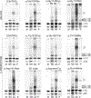
Probands with C-propeptide mutation have defective processing of procollagen. A) Pericellular processing of secreted procollagen by control cells shows an increase of mature α1(I) and α2(I) and a concomitant decrease of pro α1(I) starting at Day 2. Probands with mutations in the C-propeptide or the carboxyl end of the collagen helix have a delay in pro α1(I) processing and an increased proportion of pC-α1(I) collagen compared to control. Additionally, mature collagen is often much less abundant than control. B) Processing of the trimeric C-propeptide by BMP-1 from probands’ procollagens compared to controls (based on Table 3; BMP-1 alone). BMP-1 can only fully cleave 30–40% of the trimeric C-propeptide from probands’ procollagens compared to cleavage of control procollagens (data are normalized to 120 min incubation). C) Representative autoradiogram shows that processing of the pro-α chains from p.Gly1281Val and p.Asp1441Tyr by BMP-1 is reduced. Top and central panels, 6% gel, reduced. The C-propeptide subunits, C1 and C2, are not resolved and appear as a single band (designated C1 + C2) representing the total amount of the C-propeptide released. Bottom panel, 12% acrylamide gel, reduced where the C-propeptide subunits, C1 and C2, are resolved.
Fig. 5.
Fig. 5.
Collagens with C-propeptide mutations are deposited into the extracellular matrix and form mature cross-links. Mature cross-linked collagen from matrix deposited in culture (P) shows overmodification and backstreaking similar to secreted collagen (M). Helical mutations show an overall broadening of all matrix fractions, suggesting that a higher proportion of mutant collagen is being secreted and incorporated into the extracellular matrix. Increased levels of pC-collagen α1(I) are observed in the matrix of several probands in immature matrix fractions (AA). M: media/secreted collagen, NS: neutral salt extraction/newly incorporated collagen without cross-links, AA: acetic acid extraction/immaturely cross-linked collagen, P: pepsin extraction/fully cross-linked collagen.
Fig. 6.
Fig. 6.
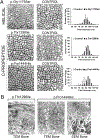
Fibrils from C-propeptide probands have round cross-sections and increased disorganization in bone. A) Dermal collagen fibrils from one helical proband (p.Gly1175Ser) and two C-propeptide probands (p.Thr1298Ile, p.Pro1444His) were examined by transmission electron microscopy (TEM) and compared with age and gender matched controls (n = 200). All probands have round fibrils, without irregular forms. p.Gly1175Ser and p.Thr1298Ile have increased fibril variability, while p.Pro1444His has a slight increase in diameter. B) Bone tissues from two C-propeptide probands were examined by TEM or scanning electron microscopy (SEM). In TEM of proband bone samples, the collagen fibrils have irregular borders and a wide distribution of fibril diameters, in contrast to proband dermal collagen fibrils. SEM of α1(I) p.Pro1444His bone tissue shows individual fibrils of highly variable sizes.

References

    1. Sillence DO, Senn A, Danks DM, Genetic heterogeneity in osteogenesis imperfecta, J. Med. Genet 16 (1979) 101–116. - PMC - PubMed
    1. Marini JC (2004) Osteogenesis imperfecta In Nelson Textbook of Pediatrics (Behrman RE, Kliegman RM and Jensen HB, eds.). pp. 2336–2338, W.B. Saunders, Philadelphia.
    1. Lees JF, Tasab M, Bulleid NJ, Identification of the molecular recognition sequence which determines the type-specific assembly of procollagen, EMBO J. 16 (1997) 908–916. - PMC - PubMed
    1. Sharma U, Carrique L, Vadon-Le Goff S, Mariano N, Georges RN, Delolme F, Koivunen P, Myllyharju J, Moali C, Aghajari N, Hulmes DJS, Structural basis of homoand heterotrimerization of collagen I, Nat. Commun 8 (2017) 14671. - PMC - PubMed
    1. DiChiara AS, Li RC, Suen PH, Hosseini AS, Taylor RJ, Weickhardt AF, Malhotra D, McCaslin DR, Shoulders MD, A cysteine-based molecular code informs collagen C-propeptide assembly, Nat. Commun 9 (2018) 4206. - PMC - PubMed

Publication types

LinkOut - more resources