Review
. 2019 Aug:266:54-65.
doi: 10.1016/j.resp.2019.04.014. Epub 2019 May 3.

Respiratory frequency plasticity during development

Affiliations
Review

Respiratory frequency plasticity during development

Stephen M Johnson et al. Respir Physiol Neurobiol. 2019 Aug.

Abstract

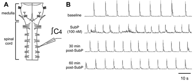
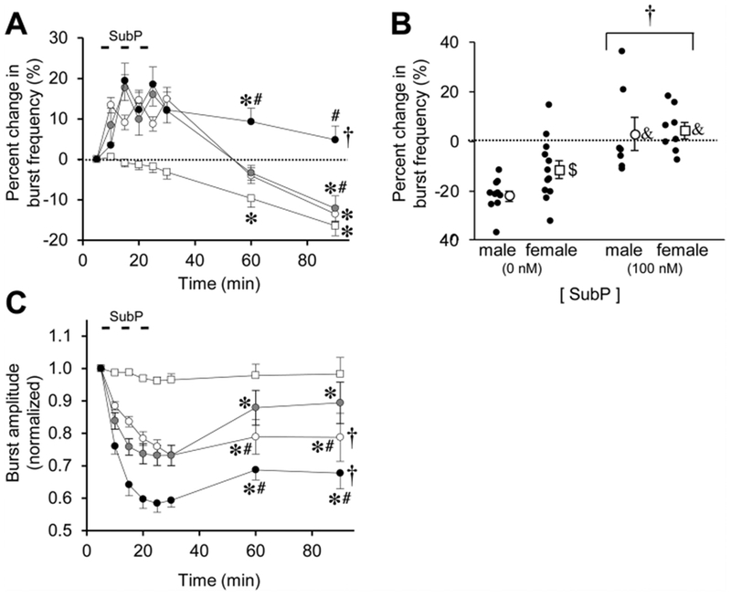
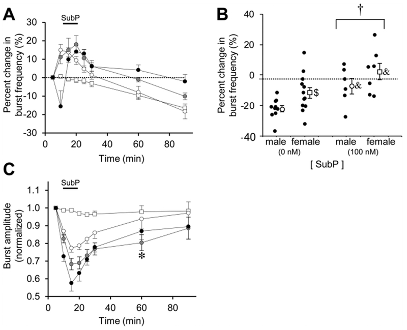
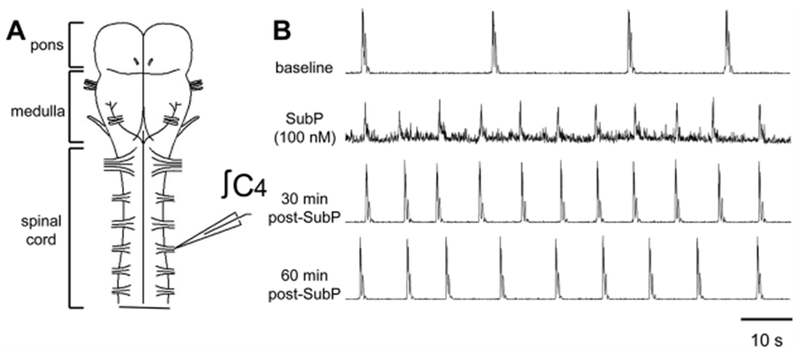
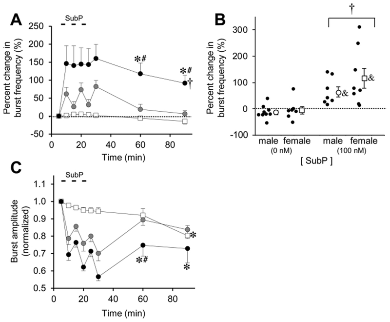
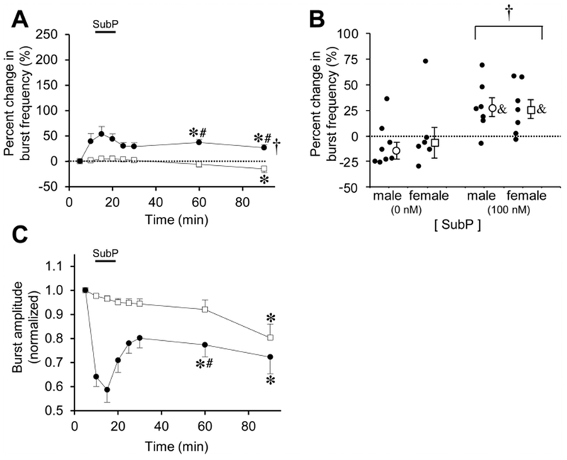
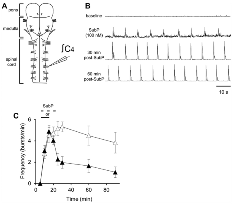
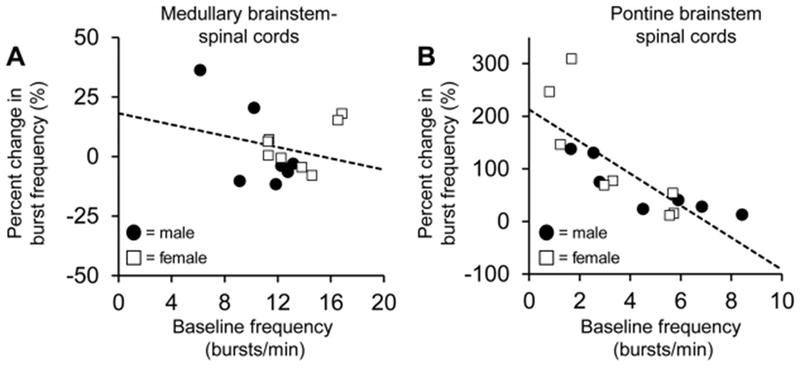
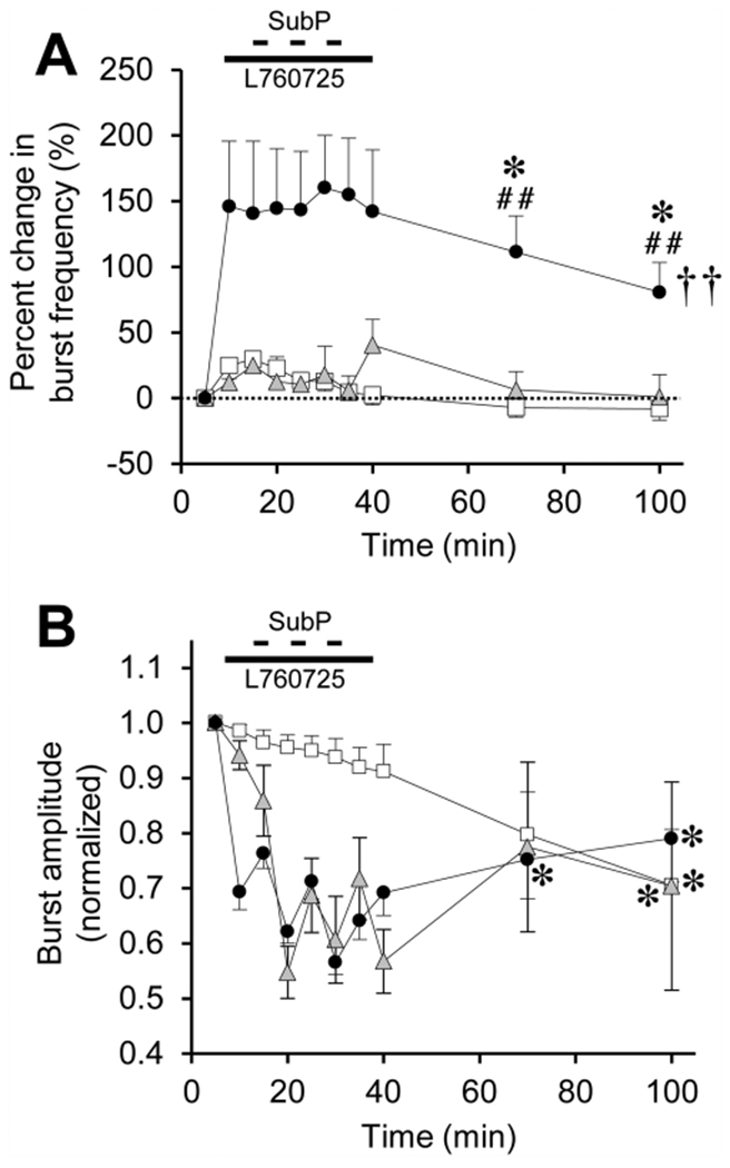
Respiratory frequency plasticity is a long-lasting increase in breathing frequency due to a perturbation. Mechanisms underlying respiratory frequency are poorly understood, and there is little evidence of frequency plasticity in neonates. This hybrid review/research article discusses available literature regarding frequency plasticity and highlights potential research opportunities. Also, we include data demonstrating a model of frequency plasticity using isolated neonatal rat brainstem-spinal cord preparations. Specifically, substance P (SubP) application induced a long-lasting (>60 min) increase in spontaneous respiratory motor burst frequency, particularly in brainstem-spinal cords with the pons attached; there were no male/female differences. SubP-induced frequency plasticity is dependent on the application pattern, such that intermittent (rather than sustained) SubP applications induce more frequency plasticity. SubP-induced frequency plasticity was blocked by a neurokinin-1 receptor antagonist. Thus, the newborn rat respiratory control system has the capacity to express frequency plasticity. Identifying mechanisms that induce frequency plasticity may lead to novel methods to safely treat breathing disorders in premature and newborn infants.

Keywords: Brain stem; Development; Frequency plasticity; Neonatal; Neuroplasticity; Respiratory; Spinal cord; Substance P.


Figures

Fig. 1.
Fig. 1.. SubP-induced changes in respiratory motor bursts produced by medullary neonatal rat brainstem-spinal cords.
(A) Drawing of isolated neonatal rat medullary brainstem-spinal cord with a suction electrode attached to the cervical spinal C4 ventral root. (B) Voltage traces of respiratory-related motor output are shown at baseline (top trace), during intermittent 100 nM SubP application (2nd trace), and at 30 and 60 min post-drug application (3rd and 4th traces, respectively). Intermittent 100 nM SubP application induced a long-lasting increase in respiratory burst frequency.
Fig. 2.
Fig. 2.. Long-lasting effects of intermittent SubP application on burst frequency and amplitude in medullary brainstem-spinal cords.
(A) The time course of burst frequency effects are shown for intermittent SubP application (3-min application/7-min washout, ×3) at 1 nM (white circles), 10 nM (gray circles), and 100 nM (black circles). Data from time control experiments are shown (white squares). Intermittent SubP applications induces frequency plasticity at 100 nM, but not at 1 or 10 nM. (B) Mean percent change in burst frequency at the 90-min time point is shown for males (white circles) and females (white squares) following 0 nM SubP (time controls) and 100 nM SubP intermittent applications. Individual data points in each group are shown as black circles. There were no sex-dependent differences in frequency plasticity, but the percent change in burst frequency was decreased in males compared to females in the time controls. (C) The time course of burst amplitude effects are shown for intermittent SubP applications. There were long-lasting decreases in burst amplitude following intermittent 100 and 1 nM SubP applications. For panels (A) and (B), statistics symbols are as follows: † = significant drug-dependent effect; * = different from baseline; # = different from time controls at that time point. For panel (B), † = significant drug-dependent effect with males and females combined; & = different from the time control for that sex; $ = difference between males and females for that [SubP].
Fig. 3.
Fig. 3.. Long-lasting effects of sustained SubP applications in medullary brainstem-spinal cords.
(A) The time course of burst frequency effects are shown for a sustained SubP application (one 9-min application) at 1 nM (white circles), 10 nM (gray circles), and 100 nM (black circles). Data from time control experiments are shown (white squares). There was no frequency plasticity observed following sustained SubP applications. (B) Mean percent change in burst frequency at the 90-min time point is shown for males (white circles) and females (white squares) following 0 nM SubP (time controls) and 100 nM SubP sustained applications. Individual data points in each group are shown as black circles. When comparing data only at the 90-min time point, frequency plasticity was expressed and there was a significant drug-dependent effect. There were no sex-dependent differences in frequency plasticity, but the percent change in burst frequency was decreased in males compared to females for time controls (as shown in Fig. 2B). (C) The time course of burst amplitude effects are shown for intermittent SubP applications. There were no long-lasting decreases in burst amplitude with sustained SubP applications. Statistics symbols as in Fig. 2 legend.
Fig. 4.
Fig. 4.. SubP-induced changes in respiratory motor bursts produced by pontine neonatal rat brainstem-spinal cords.
(A) Drawing of isolated neonatal rat pontine brainstem-spinal cord with a suction electrode attached to the cervical spinal C4 ventral root. (B) Voltage traces of respiratory-related motor output are shown at baseline (top trace), during intermittent 100 nM SubP application (2nd trace), and at 30 and 60 min post-drug application (3rd and 4th traces, respectively). Intermittent 100 nM SubP application induced a long-lasting increase in respiratory burst frequency.
Fig. 5.
Fig. 5.. Long-lasting effects of intermittent SubP application on burst frequency and amplitude in pontine brainstem-spinal cords.
(A) The time course of burst frequency effects are shown for intermittent SubP application (3-min application/7-min washout, ×3) at 10 nM (gray circles) and 100 nM (black circles). Data from time control experiments are shown (white squares). Intermittent SubP applications induced frequency plasticity at 100 nM, but not at 10 nM. (B) Mean percent change in burst frequency at the 90-min time point is shown for males (white circles) and females (white squares) following 0 nM SubP (time controls) and 100 nM SubP intermittent applications. Individual data points in each group are shown as black circles. There were no sex-dependent differences in frequency plasticity. (C) The time course of burst amplitude effects are shown for intermittent SubP applications. There were no long-lasting decreases in burst amplitude following intermittent 100 and 10 nM SubP applications compared to time controls. Statistics symbols as in Fig. 2 legend.
Fig. 6.
Fig. 6.. Long-lasting effects of sustained SubP applications in pontine brainstem-spinal cords.
(A) The time course of burst frequency effects are shown for a sustained SubP application (one 9-min application) at 100 nM (black circles). Data from time control experiments are shown (white squares). Frequency plasticity was observed following sustained SubP applications, but a much lower amount compared to intermittent 100 nM SubP applications. (B) Mean percent change in burst frequency at the 90-min time point is shown for males (white circles) and females (white squares) following 0 nM SubP (time controls) and 100 nM SubP sustained applications. Individual data points in each group are shown as black circles. When comparing data only at the 90-min time point, frequency plasticity was expressed and there was a significant drug-dependent effect. There were no sex-dependent differences in frequency plasticity. (C) There were no long-lasting decreases in burst amplitude with sustained SubP applications compared to time controls. Statistics symbols as in Fig. 2 legend.
Fig. 7.
Fig. 7.. SubP induced respiratory motor in silent pontine neonatal rat brainstem-spinal cords.
(A) Drawing of isolated neonatal rat pontine brainstem-spinal cord with a suction electrode attached to the cervical spinal C4 ventral root. (B) Voltage traces of respiratory-related motor output are shown at baseline that was silent (top trace). During intermittent 100 nM SubP application (2nd trace), respiratory motor bursts were induced, and the preparation continued to produce motor bursts for the next 30 and 60 min post-drug application (3rd and 4th traces, respectively). (C) Burst frequency in these preparations following intermittent 100 nM SubP applications (white triangles) and sustained 100 nM SubP applications (black triangles). Intermittent SubP applications to silent pontine brainstem-spinal cords tended to induce respiratory motor bursts at a higher frequency for 60 min post-drug application.
Fig. 8.
Fig. 8.. The magnitude of SubP-induced frequency plasticity is indirectly correlated with baseline burst frequency in pontine (B), but not medullary brainstem-spinal cords (A).
Individual data points are shown for males (black circles) and females (white squares). Dotted lines indicate the line of regression calculated for each data set.
Fig. 9.
Fig. 9.. SubP-induced frequency plasticity in pontine brainstem-spinal cords is abolished by a neurokinin-1 (NK1) receptor antagonist drug (L760735, 10 μM).
(A) The time course of burst frequency effects are shown for intermittent SubP applications alone (3-min application/7-min washout, ×3) at 100 nM (black circles; same data as in Fig. 5A), and intermittent SubP applications with L760735 pretreatment (gray triangles). Data from time control experiments are shown (white squares; same data as in Fig. 5A). No frequency plasticity was observed with L760735 treatment. (B) L760735 did not block the acute SubP-induced decrease in burst amplitude, and did not alter burst amplitude with respect to intermittent SubP applications only, or time control experiments. Statistics symbols as in Fig. 2 legend.

References

    1. Bach KB, Mitchell GS, 1998. Hypercapnia-induced long-term depression of respiratory activity requires alpha2-adrenergic receptors. J. Appl. Physiol 84, 2099–2105. - PubMed
    1. Bairam A, Laflamme N, Drolet C, Piedboeuf B, Shah PS, Kinkead R, 2018. Sex-based differences in apnoea of prematurity: a retrospective cohort study. Exp. Physiol 103, 1403–1411. - PubMed
    1. Baker TL, Mitchell GS, 2000. Episodic but not continuous hypoxia elicits long-term facilitation of phrenic motor output in rats. J. Physiol 529, 215–219. - PMC - PubMed
    1. Baker-Herman TL, Mitchell GS, 2008. Determinants of frequency long-term facilitation following acute intermittent hypoxia in vagotomized rats. Respir. Physiol. Neurobiol 162, 8–17. - PMC - PubMed
    1. Bavis RW, MacFarlane PM, 2017. Developmental plasticity in the neural control of breathing. Exp. Neurol 287, 176–191. - PubMed

Publication types