. 2019 Jun 4;91(11):7403-7410.
doi: 10.1021/acs.analchem.9b01351. Epub 2019 May 13.

Quantitation of Apurinic/Apyrimidinic Sites in Isolated DNA and in Mammalian Tissue with a Reduced Level of Artifacts

Affiliations

Quantitation of Apurinic/Apyrimidinic Sites in Isolated DNA and in Mammalian Tissue with a Reduced Level of Artifacts

Haoqing Chen et al. Anal Chem. .

Abstract

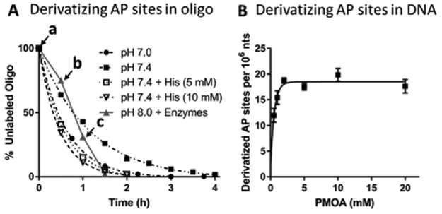
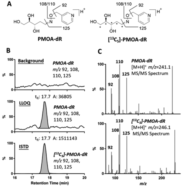
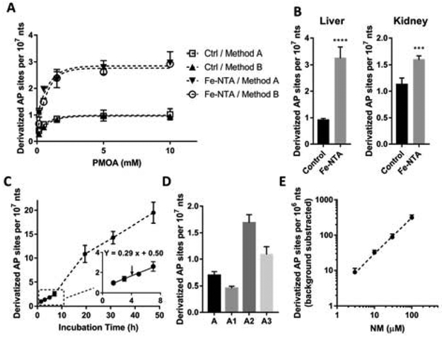
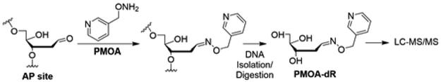
The apurinic/apyrimidinic (AP) site is a common lesion of DNA damage. The levels of AP sites reported in the literature cover a wide range, which is primarily due to the artifactual generation or loss of AP sites during processing of the DNA. Herein, we have developed a method for quantitating AP sites with a largely reduced level of artifacts by derivatizing AP sites before DNA isolation. A rapid digestion of nuclear protein was performed to minimize enzymatic DNA repair, followed by direct derivatization of AP sites in the nuclear lysate with O-(pyridin-3-yl-methyl)hydroxylamine, yielding an oxime derivative that is stable through the subsequent DNA processing steps. Quantitation was done using highly selective and sensitive liquid chromatography-tandem mass spectrometry, with a limit of quantitation at 2.2 lesions per 108 nucleotides (nts, 0.9 fmol on column). The method was applied in vivo to measure AP sites in rats undergoing oxidative stress [liver, 3.31 ± 0.47/107 nts (dosed) vs 0.91 ± 0.06/107 nts (control); kidney, 1.60 ± 0.07/107 nts (dosed) vs 1.13 ± 0.12/107 nts (control)]. The basal AP level was significantly lower than literature values. The method was also used to measure AP sites induced by the chemotherapeutic nitrogen mustard in vitro.


Figures

Figure 1.
Figure 1.
(A) Derivatization of AP sites in double-stranded oligonucleotide with 5 mM PMOA at 37 °C, with different buffers and catalysts. The percent unreacted oligonucleotide vs. time is reported. Points on the reaction rate curve at pH 8.0 containing sequential addition of enzymes employed during lysis of the nuclear pellet: a. RNase A & RNase T1 added. b. Proteinase K added, c. Puregene PP solution added, or reactions conducted at pH 7.0 or pH 7.4 without or with histidine. (B) Derivatization of AP sites in CT DNA with various concentrations of PMOA. Reaction condition: pH 7.4, 37 °C, 1.5 h. Each value represents the mean ± SD, n = 3.
Figure 2.
Figure 2.
(A) Proposed MS/MS fragmentation of PMOA-dR ([M+H]+ m/z 241.1) and [13C5]-PMOA-dR ([M+H]+ m/z 246.1). (B) Extracted ion chromatogram (EIC) of PMOA-dR (at background level normalized against the base peak in LLOQ, and at LLOQ) and [13C5]-PMOA-dR (1.1 per 106 nts) in DNA digestion matrix and (C) PMOA-dR and [13C5]-PMOA-dR MS/MS spectra. The * represent the sites of the 13C atoms in the internal standard.
Figure 3.
Figure 3.
(A) Derivatization of AP sites in rat liver nuclei. Rats were sacrificed 6 h after intraperitoneal injection with Fe-NTA (15 mg Fe/kg) and vehicle (Ctrl group). AP sites were derivatized by the nuclear lysate-derivatization method (method A) or nucleic acid-derivatization method (method B). Each value represents the mean ± SD, n = 3. (B) AP site level in rat livers and kidneys after administration of Fe-NTA (grey bar) or vehicle (black bar). n = 4. Two-tailed t test p < 0.0001 (****), p < 0.001 (***). (C) Spontaneous depurination rate measured in untreated rat liver nuclei lysate (n=3). (D) AP site level in DNA isolated/derivatized by different methods. A. After nuclear isolation, conduct proteolysis for 1.5 h and then AP site derivatization for 1.5 h. A1: After nuclear isolation, conduct AP site derivatization and proteolysis together for 1.5 h. A2: Treat nuclei with RNases for 1.5 h, proteinase K for 2 h, and precipitate excess proteins. Precipitate DNA with salt/isopropanol and immediately derivatize the isolated DNA. A3: Unlike method A2, DNA pellet was precipitated under −20 °C overnight before derivatization. n = 4. (E) Isolated rat liver DNA was incubated with NM at 37 °C for 1 h, followed by neutral hydrolysis for 5 h (pH 7.4). n = 3. The background level of AP sites by neutral hydrolysis (6.5 ± 1.5/107 nts) was subtracted.
Scheme 1.
Scheme 1.
Quantitation of AP sites by PMOA derivatizing method.

References

    1. Kim Y-J; Wilson DM Curr. Mol. Pharmacol 2012, 5, 3–13. - PMC - PubMed
    1. Lindahl T In Prog. Nucleic Acid Res. Mol. Biol, Cohn WE, Ed.; Academic Press, 1979, pp 135–192. - PubMed
    1. Krokan HE; Standal R; Slupphaug G Biochem. J 1997, 325, 1–16. - PMC - PubMed
    1. De Bont R; van Larebeke N Mutagenesis 2004, 19, 169–185. - PubMed
    1. Nakamura J; Walker VE; Upton PB; Chiang S-Y; Kow YW; Swenberg JA Cancer Res. 1998, 58, 222–225. - PubMed

Publication types

Substances