. 2019 Jul 18;26(7):960-969.e4.
doi: 10.1016/j.chembiol.2019.03.014. Epub 2019 May 2.

Allosteric Regulation of Oligomerization by a B12 Trafficking G-Protein Is Corrupted in Methylmalonic Aciduria

Affiliations

Allosteric Regulation of Oligomerization by a B12 Trafficking G-Protein Is Corrupted in Methylmalonic Aciduria

Markus Ruetz et al. Cell Chem Biol. .

Abstract

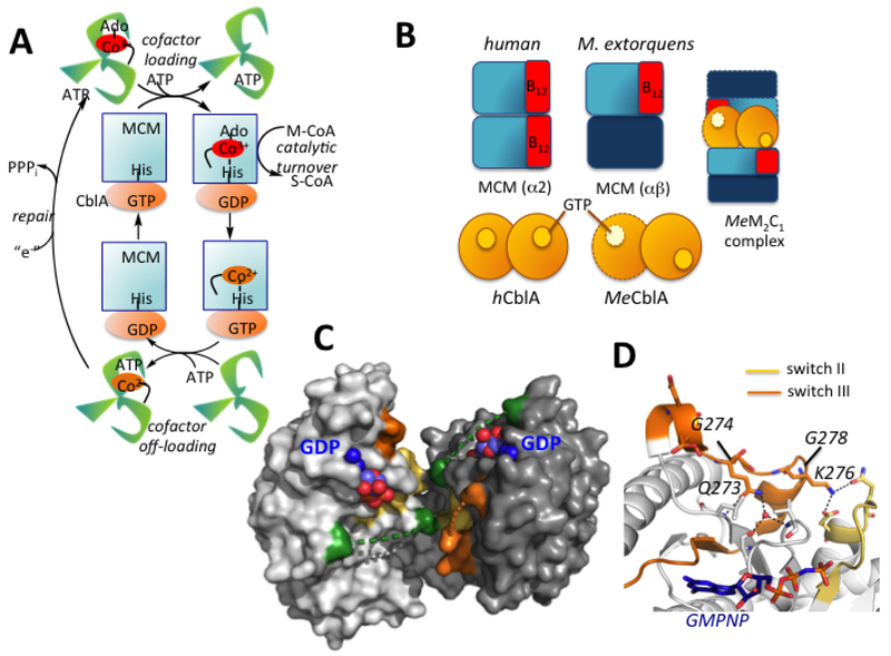
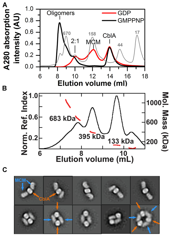
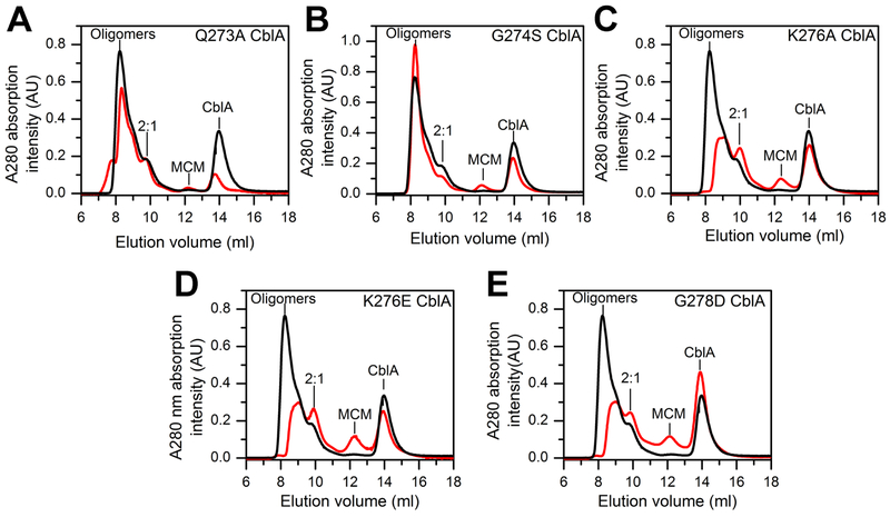
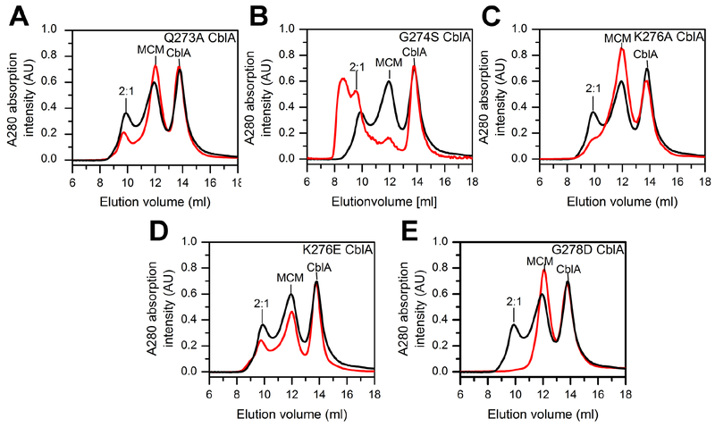
Allosteric regulation of methylmalonyl-CoA mutase (MCM) by the G-protein chaperone CblA is transduced via three "switch" elements that gate the movement of the B12 cofactor to and from MCM. Mutations in CblA and MCM cause hereditary methylmalonic aciduria. Unlike the bacterial orthologs used previously to model disease-causing mutations, human MCM and CblA exhibit a complex pattern of regulation that involves interconverting oligomers, which are differentially sensitive to the presence of GTP versus GDP. Patient mutations in the switch III region of CblA perturb the nucleotide-sensitive distribution of the oligomeric complexes with MCM, leading to loss of regulated movement of B12 to and/or from MCM and explain the molecular mechanism of the resulting disease.

Keywords: G-protein; GTPase; MMAA; cblA; cobalamin; cofactor; metal trafficking; metalloprotein; trafficking; vitamin B(12).


Conflict of interest statement

Declaration of Interest

The authors declare that no competing financial interests exist.

Figures

Figure 1.
Figure 1.. Chaperone functions and structure of human CblA.
(A) AdoCbl is loaded onto MCM from ATR in a process gated by the GTPase activity of CblA. MCM has GAP function and enhances the GTPase activity of CblA when the two proteins form a complex. Holo- MCM catalyzes the isomerization of methylmalonyl-CoA (M-CoA) to succinyl-CoA (S-CoA). When MCM is inactivated, the GTPase activity of CblA is used to off-load cob(II)alamin to the ATR active site. ATR completes the repairs process, reforming AdoCbl. (B) Cartoon showing differences between human and M. extorquens MCM and CblA and the M extorquens M2C1 complex that was previously characterized. (C) Structure of human CblA (PDB: 2WWW) with GDP bound. Switch I (green), II (yellow) and III (orange) loops are highlighted. Dashed lines represent disordered regions in switch I and III loops. (D) Since switch III residues 268–274 in human CblA are disordered, a close-up of the homologous region in MeMeaB (PDB: 4YJB) is shown with the location of residues (human numbering) mutated in this study.
Figure 2.
Figure 2.. Oligomeric states of MCM-wild-type CblA complexes.
(A) Distribution of complexes formed upon mixing MCM (25 μM) and CblA A (50 μM) in the presence of GMPPNP or GDP (500 μM each). Positions of the calibration standards are indicated in the light gray trace. (B) SEC-MALS analysis of a mixture of MCM (25 μM) and wild-type CblA (25 μM) in the presence of GMPPNP (250 μM). The black trace represents the normalized refractive index (left axis). The red line indicates the molecular weight as detected by MALS (right axis). (C) 2D class averages of the 395 kDa peak shows heterogeneity of oligomeric forms. The orange and blue arrows denote CblA and MCM, respectively.
Figure 3.
Figure 3.
Oligomeric distribution of MCM-CblA complexes in the presence of GMPPNP. MCM (25 μM) and wild-type or mutant CblA (50 μM each) were incubated with GMPPNP (500 μM) for 4 min at 4°C. The black trace in each panel corresponds to the elution profile with wild-type CblA and the red trace with the following CblA mutants: (A) Q273A (B) G274S (C) K276A, (D) K276E, and (E) G278D. The M2C1 complex is simply denoted as 2:1 in the panels. The data are representative of two independent experiments.
Figure 4.
Figure 4.
Oligomeric distribution of MCM-CblA complexes in the presence of GDP. MCM (25 μM) and wild-type or mutant CblA (50 μM each) were incubated with GDP (500 μM) for 4 min at 4°C. In each panel, the black trace corresponds to the elution profile with wild-type CblA and the red trace in the presence of the following CblA mutant: (A) Q273A, (B) G274S, (C) K276A, (D) K276E, and (E) G278D. The data are representative of two independent experiments.
Figure 5.
Figure 5.. Switch III mutations affect MCM-CblA oligomeric forms.
(A) MCM (25 μM) was mixed with wild-type (50 μM, black line), G274S (red line) or K276E CblA (blue line) CblA and GMPPCP (500 μM) and separated by analytical gel filtration. The fractions indicated by the numbers 1–3 (at 8.2, 9.0 and 9.8 ml) were diluted to a 10 μg/ml protein concentration and transferred onto the EM grid. (B) Selected reference-free 2D class averages showing the circular organization of MCM-wild-type CblA•GMPPCP complexes in fractions 2 and 3. Box length, 50.4 nm. The number of particles in each average shown in right corner. The lower resolution of the class averages in fraction 2 is due to the exhibit higher flexibility of the particles. (C) The number of circular particles averaged for 100 negative stained images for each MCM-CblA complex (fraction 3).
Figure 6.
Figure 6.. Model for AdoCbl loading and offloading in human.
(A) Transfer of AdoCbl (15 μM) from ATR (15 μM trimer) to MCM (15 μM) in the presence of wild-type CblA (45 μM) and 1 mM GDP or GMPPCP (inset). The initial and final spectra (30 min) are in black and red, respectively. (B) Transfer of cob(II)alamin (15 μM) bound to MCM (9 μM, black trace) in the presence of CblA (30 μM) following addition of ATR (15 μM), ATP (5 mM) and 1 mM GTP or GMPPCP (inset). The final spectrum after 15 min is in red. (C) Model for cofactor loading/off-loading highlighting a central role for the GTPase activity of CblA. Resolution of the higher order oligomers (left) to a “loading ready” M2C1 complex is driven by GTP hydrolysis, which following AdoCbl transfer separates into the individual proteins. During catalysis, occasional inactivation of MCM leads to the loss of deoxyadenosine and accumulation of cob(II)alamin, which is off-loaded in the presence of CblA in a GTPase- dependent step. For simplicity, the offloading complex is shown as M2C1 although it is currently unclear whether it can also occur from the M1C1 or the higher order complexes.

References

    1. Ando N, Brignole EJ, Zimanyi CM, Funk MA, Yokoyama K, Asturias FJ, Stubbe J, and Drennan CL (2011). Structural interconversions modulate activity of Escherichia coli ribonucleotide reductase. Proc Natl Acad Sci U S A 108, 21046–21051. - PMC - PubMed
    1. Banerjee R (2003). Radical carbon skeleton rearrangements: catalysis by coenzyme B12-dependent mutases. Chem Rev 103, 2083–2094. - PubMed
    1. Banerjee R, Gherasim C, and Padovani D (2009). The Tinker, Tailor, Soldier in intracellular B12 Trafficking. Cur Op Chem Biol 13, 484–491. - PMC - PubMed
    1. Breinig S, Kervinen J, Stith L, Wasson AS, Fairman R, Wlodawer A, Zdanov A, and Jaffe EK (2003). Control of tetrapyrrole biosynthesis by alternate quaternary forms of porphobilinogen synthase. Nat Struct Biol 10, 757–763. - PubMed
    1. Campanello GC, Lofgren M, Yokom AL, Southworth DR, and Banerjee R (2017). Switch I-dependent allosteric signaling in a G-protein chaperone-B12 enzyme complex. J Biol Chem 292, 17617–17625. - PMC - PubMed

Publication types

MeSH terms

Supplementary concepts