. 2019 May 7;8(9):e011630.
doi: 10.1161/JAHA.118.011630.

Arterial Stiffness Due to Carotid Calcification Disrupts Cerebral Blood Flow Regulation and Leads to Cognitive Deficits

Affiliations

Arterial Stiffness Due to Carotid Calcification Disrupts Cerebral Blood Flow Regulation and Leads to Cognitive Deficits

Gervais Muhire et al. J Am Heart Assoc. .

Abstract

Background Arterial stiffness is associated with cognitive decline and dementia; however, the precise mechanisms by which it affects the brain remain unclear. Methods and Results Using a mouse model based on carotid calcification this study characterized mechanisms that could contribute to brain degeneration due to arterial stiffness. At 2 weeks postcalcification, carotid stiffness attenuated resting cerebral blood flow in several brain regions including the perirhinal/entorhinal cortex, hippocampus, and thalamus, determined by autoradiography ( P<0.05). Carotid calcification impaired cerebral autoregulation and diminished cerebral blood flow responses to neuronal activity and to acetylcholine, examined by laser Doppler flowmetry ( P<0.05, P<0.01). Carotid stiffness significantly affected spatial memory at 3 weeks ( P<0.05), but not at 2 weeks, suggesting that cerebrovascular impairments precede cognitive dysfunction. In line with the endothelial deficits, carotid stiffness led to increased blood-brain barrier permeability in the hippocampus ( P<0.01). This region also exhibited reductions in vessel number containing collagen IV ( P<0.01), as did the somatosensory cortex ( P<0.05). No evidence of cerebral microhemorrhages was present. Carotid stiffness did not affect the production of mouse amyloid-β (Aβ) or tau phosphorylation, although it led to a modest increase in the Aβ40/Aβ42 ratio in frontal cortex ( P<0.01). Conclusions These findings suggest that carotid stiffness alters brain microcirculation and increases blood-brain barrier permeability associated with cognitive impairments. Therefore, arterial stiffness should be considered a relevant target to protect the brain and prevent cognitive dysfunctions.

Keywords: arterial stiffness; blood‐brain barrier; carotid calcification; cerebral blood flow; cognitive impairment.


Figures

Figure 1
Figure 1
Effect of carotid calcification on spatial learning and memory. Cognitive abilities were examined with the Morris Water Maze test (2 and 3 weeks after application of CaCl2 or NaCl) (A and C). The test consisted of 5 training days (D1–D5) during which mice were allowed three 60 seconds trials per day to find the escape platform. Each trial was 20 minutes apart. The area under the learning curve (AUC) and the average latency across the training (B and D) were calculated to compare learning performance. Graphs represent mean±SD. Data are displayed in box‐and‐whisker plots, where the median is represented by the horizontal line, and the 10th and 90th percentiles extend from the extremes of the box. For the probe test (day 6), the platform was removed and animals were given two 30 seconds trials separated 1 hour apart, to assess (E and H) time spent in the target quadrant (the place where the platform was originally located), (F and I) number of target crossings and (G and J) latency to reach the target zone. Data were analyzed with ANOVA for factorial design with repeated measures followed by a Fisher's LSD posttest for multiple group comparisons for the learning curves; unpaired Student t‐test was used for all other 2‐group comparisons; n=8 to 9 mice/group (2 weeks) and n=9 to 13 mice/group (3 weeks). *P<0.05; TQ indicates target quadrant.
Figure 2
Figure 2
Effect of carotid calcification on resting cerebral blood flow (CBF). Resting CBF was measured by autoradiography using [14C]iodoantipyrine as a diffusible tracer in awake mice, 2 weeks after application of CaCl2 or NaCl. CBF was quantified in the following regions from the right hemisphere: visual and somatosensory cortices (VSCx), perirhinal and entorhinal cortices (PECx), cornu ammonis area 1 (CA1), cornu ammonis area 2‐3 (CA2‐3), dentate gyrus (DG), thalamus (THA), and corpus callosum (CC). Data are displayed in box‐and‐whisker plots, where the median is represented by the horizontal line and the 10th and 90th percentiles extend from the extremes of the box. *P<0.05; unpaired Student t‐test per independent brain region (results presented in a single graph); n=5 to 7 mice/group. CBF is expressed as mL/100 g/min. Representative blood flow autoradiograms for control (upper panel) and CaCl2 mice (lower panel) are shown with a color scale.
Figure 3
Figure 3
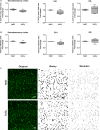
Effect of carotid calcification on number of cerebral vessel containing collagen type IV. Semiquantitative analysis of microvessel number per field by examination of (A) collagen IV–positive area percentage and (B) vascular skeleton area in the somatosensory cortex and hippocampus (cornu ammonis area 1 [CA1] and dentate gyrus [DG]) from the right hemisphere. Data are displayed in box‐and‐whisker plots, where the median is represented by the horizontal line, and the 10th and 90th percentiles extend from the extremes of the box. *P<0.05, **P<0.01, ***P<0.0001; unpaired Student t‐test, n=5 to 6 mice/group. C, Representative micrographs of collagen IV immunostaining, binary transformation, and skeletonization for CA1 are shown. Scale bar=50 μm. Analysis was done 3 weeks after surgery.
Figure 4
Figure 4
Effect of carotid calcification on cerebrovascular responses. A, Analysis of cerebral autoregulation depicting relationship between mean arterial pressure and cerebral blood flow (CBF). Graph represents mean±SD. Data are displayed in box‐and‐whisker plots, where the median is represented by the horizontal line, and the 10th and 90th percentiles extend from the extremes of the box. ***P<0.001, linear regression of slopes between 70 and 130 mmHg was used; n=5 to 9 mice/group. CBF responses to whisker stimulation (B), to the endothelium‐dependent vasodilator, acetylcholine (C), and to the endothelium‐independent vasodilator, sodium nitroprussiate (SNP; D) were evaluated. Graphs depict the percentage increase in CBF following the stimulation with respect to its initial value. *P<0.05; **P<0.01; unpaired Student t‐test; n=4 to 6 mice/group. CBF was measured on the right somatosensory cortex by laser Doppler flowmetry 2 weeks after application of CaCl2 or NaCl.
Figure 5
Figure 5
Effect of carotid calcification on blood‐brain barrier permeability. Permeability of the blood‐brain barrier was examined by sodium fluorescein (NaF) extravasation in specific brain regions cortex (A), frontal cortex (B) and hippocampus (C) from the right hemisphere 3 weeks after application of CaCl2 or NaCl. The amount of NaF in each region was calculated from a standard curve (0–5 μmol/L NaF) and normalized per gram of tissue. Results are expressed as fold change by normalizing values to the average of the control group. Data are displayed in box‐and‐whisker plots, where the median is represented by the horizontal line, and the 10th and 90th percentiles extend from the extremes of the box. **P<0.01; unpaired Student t‐test, n=7 mice/group.
Figure 6
Figure 6
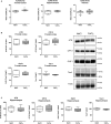
Effect of carotid calcification on amyloid‐β (Aβ) and tau hyperphosphorylation. A, The Aβ40/Aβ42 ratio was calculated from Aβ concentrations determined by ELISA in the frontal cortex and hippocampal tissue as well as in plasma. Expression of phosphorylated tau (AT8, CP13 and PHF1) and dephosphorylated tau (Tau1) in frontal cortex (B) and hippocampus (C) examined by western blotting. Representative immunoblots are shown. Data are displayed in box‐and‐whisker plots, where the median is represented by the horizontal line and th,e 10th and 90th percentiles extend from the extremes of the box. **P<0.01; unpaired Student t‐test, n=6 to 7 mice/group in frontal cortex and hippocampus, n=10 to 15 mice/group in plasma. Analysis was done 3 weeks after surgery on frozen tissue from the right hemisphere.

References

    1. Pase MP, Herbert A, Grima NA, Pipingas A, O'Rourke MF. Arterial stiffness as a cause of cognitive decline and dementia: a systematic review and meta‐analysis. Intern Med J. 2012;42:808–815. - PubMed
    1. Hughes TM, Craft S, Lopez OL. Review of “the potential role of arterial stiffness in the pathogenesis of Alzheimer's disease.” Neurodegener Dis Manag. 2015;5:121–135. - PMC - PubMed
    1. Hanon O, Haulon S, Lenoir H, Seux ML, Rigaud AS, Safar M, Girerd X, Forette F. Relationship between arterial stiffness and cognitive function in elderly subjects with complaints of memory loss. Stroke. 2005;36:2193–2197. - PubMed
    1. Meyer ML, Palta P, Tanaka H, Deal JA, Wright J, Knopman DS, Griswold ME, Mosley TH, Heiss G. Association of central arterial stiffness and pressure pulsatility with mild cognitive impairment and dementia: the Atherosclerosis Risk in Communities Study‐Neurocognitive Study (ARIC‐NCS). J Alzheimers Dis. 2017;57:195–204. - PMC - PubMed
    1. Pase MP, Beiser A, Himali JJ, Tsao C, Satizabal CL, Vasan RS, Seshadri S, Mitchell GF. Aortic stiffness and the risk of incident mild cognitive impairment and dementia. Stroke. 2016;47:2256–2261. - PMC - PubMed

Publication types

MeSH terms