. 2019 Apr 16:10:370.
doi: 10.3389/fphar.2019.00370. eCollection 2019.

Non-canonical Notch Signaling Regulates Actin Remodeling in Cell Migration by Activating PI3K/AKT/Cdc42 Pathway

Affiliations

Non-canonical Notch Signaling Regulates Actin Remodeling in Cell Migration by Activating PI3K/AKT/Cdc42 Pathway

Lei Liu et al. Front Pharmacol. .

Abstract

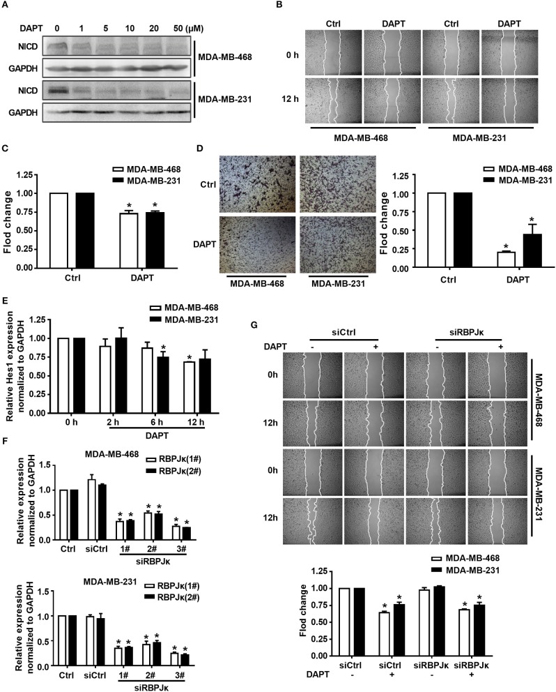
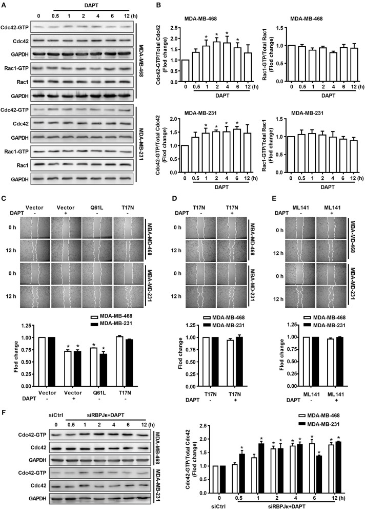
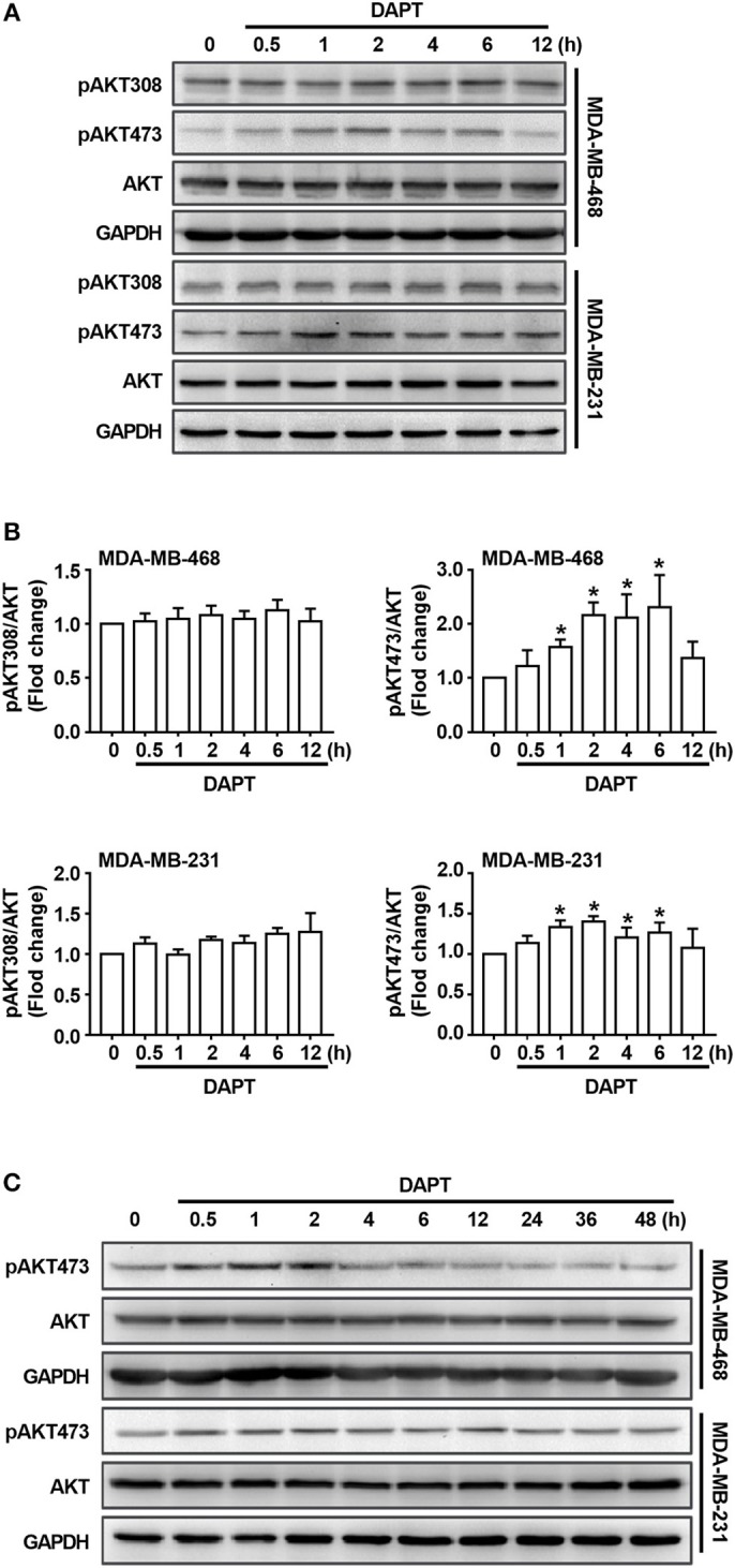
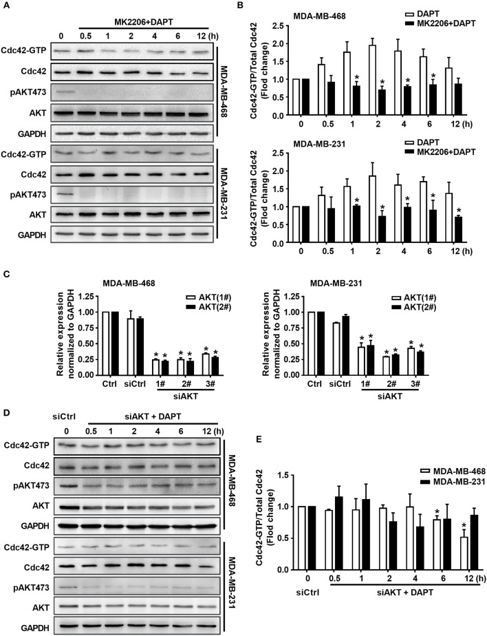
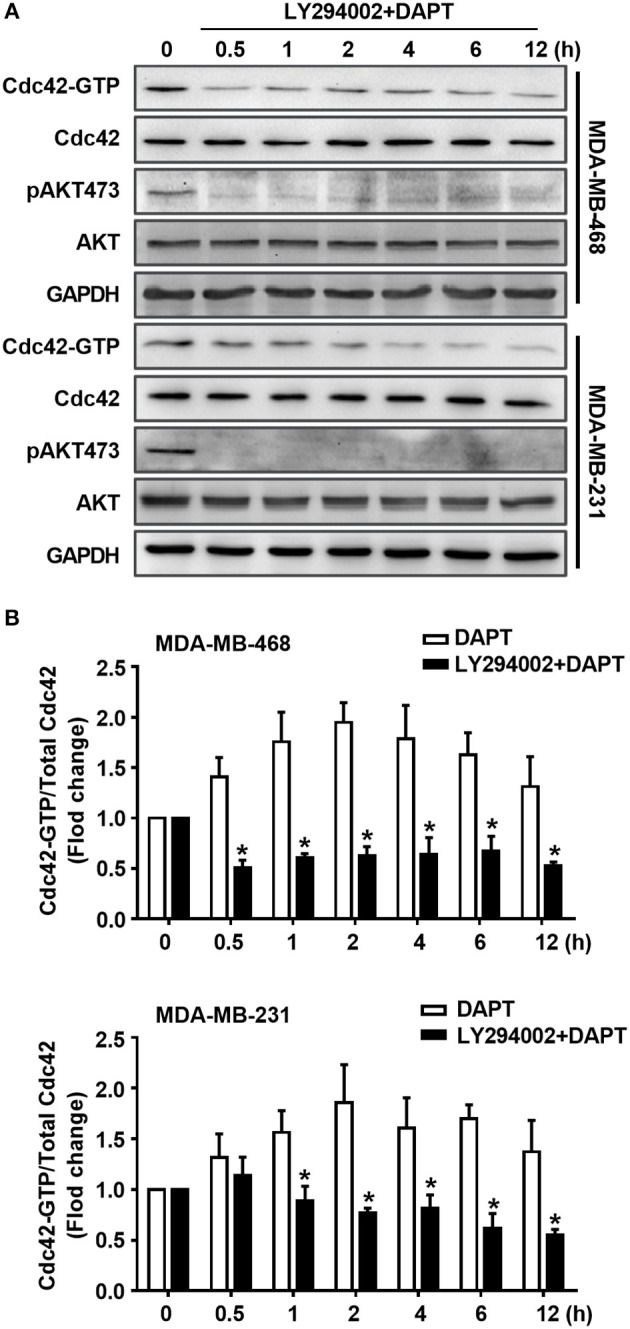
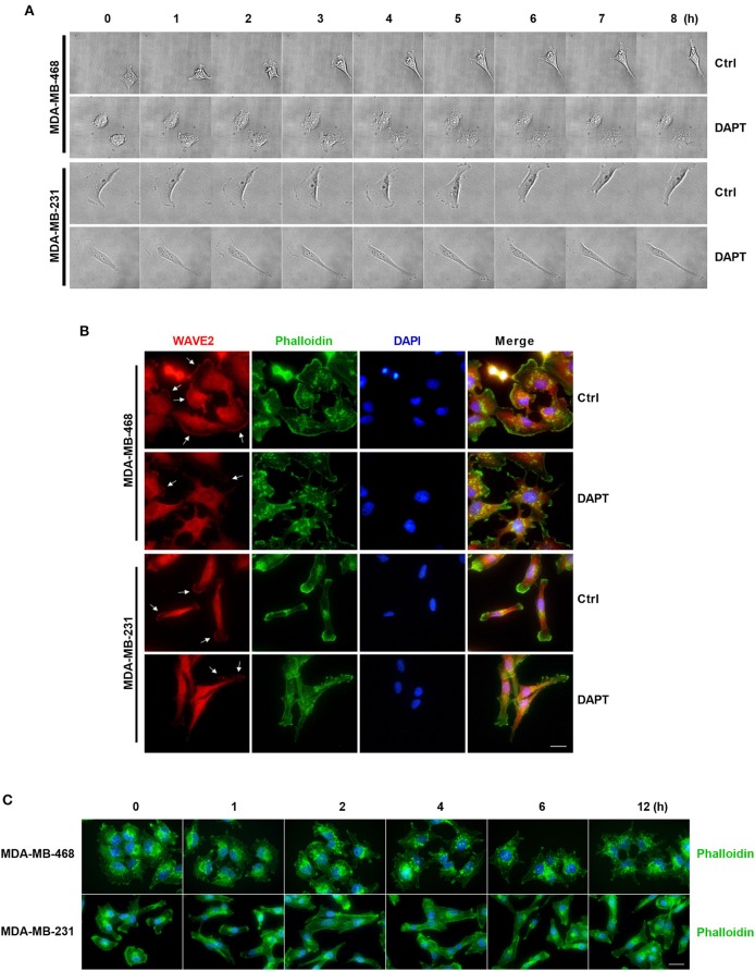
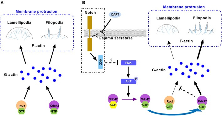
Tumor cell migration is a critical step in cancer metastasis. Over-activated Notch pathway can promote the migration of cancer cells, especially in the breast cancer. However, the underlying mechanism of non-canonical Notch signaling in modulating the migration has not yet been clearly characterized. Here we demonstrated that DAPT, a gamma secretase inhibitor, inhibited protrusion formation and cell motility, and then reduced the migration of triple-negative breast cancer cells, through increasing the activity of Cdc42 by non-canonical Notch pathway. Phosphorylation of AKT on S473 was surprisingly increased when Notch signaling was inhibited by DAPT. Inhibition of PI3K and AKT by LY294002 and MK2206, respectively, or knockdown of AKT expression by siRNA blocked DAPT-induced activation of Cdc42. Moreover, immunofluorescence staining further showed that DAPT treatment reduced the formation of lamellipodia and induced actin cytoskeleton remodeling. Taken together, these results indicated that DAPT inhibited Notch signaling and consequently activated PI3K/AKT/Cdc42 signaling by non-canonical pathway, facilitated the formation of filopodia and inhibited the assembly of lamellipodia, and finally resulted in the decrease of migration activity of breast cancer cells.

Keywords: Cdc42; PI3K/AKT; actin cytoskeleton; membrane protrusions; non-canonical Notch.


Figures

Figure 1
Figure 1
Inhibition of Notch by DAPT reduces breast cancer migration by non-canonical Notch pathway in 12 h. (A) The expression of NICD was detected in MDA-MB-468 and MDA-MB-231 cells when the cells were treated with 0–50 μM DAPT. (B) Images were taken at 0 and 12th h after wounding by an inverted microscope. (C) Relative migration rate was calculated and the migration of DAPT untreated cells at 12 h was set as 100%. *P < 0.05 DAPT treated cells at 12 h vs. DAPT untreated cells at 12 h. (D) Transwell assay was performed on control vs. 20 μM DAPT treated cells to elevate the migration of cells. Migration index of Ctrl cells at 12 h was set at 100%. *P < 0.05 DAPT-treated cells vs. Ctrl cells. (E) The effect of DAPT on the expression of Hes1 mRNA at indicated time. *P < 0.05 DAPT-treated time point vs. DAPT treated 0 h. (F) The effect of siRBPJκ on the expression of RBPJκ mRNA. *P < 0.05 cells transfected with siRBPJκ vs. cells transfected with siCtrl. (G) The effect of DAPT on the migration of cells transfected with siCtrl or siRBPJκ. *P < 0.05 DAPT-treated cells vs. DAPT-untreated cells.
Figure 2
Figure 2
The migration of breast cancer cells was inhibited by DAPT through Cdc42 pathway. (A,B) The ratio of Cdc42-GTP/Cdc42 and Rac1-GTP/Rac1 were analyzed by Pulldown assay in MDA-MB-468 and MDA-MB-231 cells, which were incubated with DAPT (20 μM) for indicated time. *P < 0.05 DAPT-treated time point vs. DAPT treated 0 h. (C) Effects of Cdc42-Q61L and Cdc42-T17N on the migration of cells. *P < 0.05 cells transfected with plasmid vs. cells transfected with vector. (D,E) Inhibition of Cdc42-T17N or ML141 (Cdc42 inhibitor, 10 μM) on the DAPT-induced migration of MDA-MB-468 and MDA-MB-231 cells. *P < 0.05 DAPT-treated cells vs. DAPT-untreated cells. (F) Analysis of the level of active Cdc42 in breast cancer cells transfected with siRBPJκ (3#) and then incubated with DAPT (20 μM) for indicated time. *P < 0.05 cells transfected with siRBPJκ vs. cells transfected with siCtrl.
Figure 3
Figure 3
DAPT increases the level of pAKT473 in breast cancer. (A) The changes of the level of T308- and S473-phosphorylated AKT in cells incubated with DAPT (20 μM) for indicated time points. (B) Relative level of T308- and S473-phosphorylated AKT in cells incubated with DAPT for indicated time points were calculated. Phosphorylated AKT level of DAPT-untreated cells was set as 100%. *P < 0.05 DAPT-treated time point vs. DAPT-treated 0 h. (C) The changes of the level of S473-phosphorylated AKT in MDA-MB-468 and MDA-MB-231 cells incubated with DAPT (0–48 h).
Figure 4
Figure 4
DAPT increases the ratio of Cdc42-GTP/Cdc42 by activating AKT. (A) The activity of Cdc42 was analyzed by Pulldown assay in MDA-MB-468 and MDA-MB-231 cells when the cells were treated with MK2206 (AKT inhibitor, 5 μM) 30 min before administration DAPT (20 μM) for indicated time points. (B) The quantification of the ratio of Cdc42-GTP/Cdc42 was performed when the cells were treated or untreated with MK2206 before administration DAPT for indicated time. *P < 0.05 MK2206-treated cells vs. MK2206-untreated cells at corresponding time point. (C) The effect of siAKT on the expression of AKT. *P < 0.05 cells transfected with siAKT vs. cells transfected with siCtrl. (D,E) The activity of Cdc42 and quantification of the ratio of Cdc42-GTP/Cdc42 in breast cancer cells transfected with siAKT (1#+2#) and then incubated with DAPT (20 μM) for indicated time points were analyzed. *P < 0.05 cells transfected with siAKT (1#+2#) vs. cells transfected with siCtrl.
Figure 5
Figure 5
DAPT activates Cdc42 through PI3K/AKT pathway in MDA-MB-231 and MDA-MB-468 cells. (A) The activity of Cdc42 was analyzed by Pulldown assay in MDA-MB-468 and MDA-MB-231 cells when the cells were treated with LY294002 (PI3K inhibitor, 10 μM) for 30 min before administration DAPT (20 μM) for indicated time. (B) The quantification of the ratio of Cdc42-GTP/Cdc42 was performed when the cells were treated or untreated with LY294002 before administration DAPT for indicated time. *P < 0.05 LY294002 treated cells vs. LY294002 untreated cells at corresponding time point.
Figure 6
Figure 6
DAPT inhibits the formation of lamellipodia and induces the F-actin remodeling during migration of breast cancer cells. (A) The changes of protrusion formation and the cell motility of DAPT-treated or DAPT-untreated breast cancer cells were observed by live cell imaging system. (B) Immunofluorescence staining showing that the distribution of WAVE2 was changed by treating breast cancer cells with DAPT. White arrows represent WAVE2 aggregation under the membrane. Scale bar, 20 μM. (C) DAPT induced the F-actin remodeling and changed cell morphology in MDA-MB-468 and MDA-MB-231 cells. Scale bar, 20 μM.
Figure 7
Figure 7
The model illustrates the mechanism for the effect of DAPT on modulating F-actin polymerization through PI3K/AKT/Cdc42 signaling by Non-canonical Notch pathway during cancer cell migration. (A) Rac1 and Cdc42 compete each other for G-actin to form different protrusive structures in cell migration. (B) DAPT activates PI3K/AKT/Cdc42 pathway through non-canonical Notch signaling. Activation of Cdc42 enhance its competition with Rac1 to bind with G-actin, which polymerizes spike-like filopodia and inhibits Rac1-based lamellipodia formation, leading to the decrease of force generation at the front of cell and the reduction of cell migration.

References

    1. Algayadh I. G., Dronamraju V., Sylvester P. W. (2016). Role of Rac1/WAVE2 signaling in mediating the inhibitory effects of gamma-tocotrienol on mammary cancer cell migration and invasion. Biol. Pharm. Bull. 39, 1974–1982. 10.1248/bpb.b16-00461 - DOI - PubMed
    1. Andersen P., Uosaki H., Shenje L. T., Kwon C. (2012). Non-canonical Notch signaling: emerging role and mechanism. Trends Cell Biol. 22, 257–265. 10.1016/j.tcb.2012.02.003 - DOI - PMC - PubMed
    1. Ayaz F., Osborne B. A. (2014). Non-canonical notch signaling in cancer and immunity. Front. Oncol. 4:345. 10.3389/fonc.2014.00345 - DOI - PMC - PubMed
    1. Bolós V., Mira E., Martínez-Poveda B., Luxán G., Cañamero M., Martínez-A C., et al. . (2013). Notch activation stimulates migration of breast cancer cells and promotes tumor growth. Breast Cancer Res. 15:R54. 10.1186/bcr3447 - DOI - PMC - PubMed
    1. Brzozowa-Zasada M., Piecuch A., Dittfeld A., Mielanczyk Ł, Michalski M., Wyrobiec G., et al. . (2016). Notch signalling pathway as an oncogenic factor involved in cancer development. Contemp. Oncol. 20, 267–272. 10.5114/wo.2016.61845 - DOI - PMC - PubMed

LinkOut - more resources