Review
. 2019 Apr 18:10:441.
doi: 10.3389/fpls.2019.00441. eCollection 2019.

Evolution of Cell Wall Polymers in Tip-Growing Land Plant Gametophytes: Composition, Distribution, Functional Aspects and Their Remodeling

Affiliations
Review

Evolution of Cell Wall Polymers in Tip-Growing Land Plant Gametophytes: Composition, Distribution, Functional Aspects and Their Remodeling

Jérémy Dehors et al. Front Plant Sci. .

Abstract

During evolution of land plants, the first colonizing species presented leafy-dominant gametophytes, found in non-vascular plants (bryophytes). Today, bryophytes include liverworts, mosses, and hornworts. In the first seedless vascular plants (lycophytes), the sporophytic stage of life started to be predominant. In the seed producing plants, gymnosperms and angiosperms , the gametophytic stage is restricted to reproduction. In mosses and ferns, the haploid spores germinate and form a protonema, which develops into a leafy gametophyte producing rhizoids for anchorage, water and nutrient uptakes. The basal gymnosperms (cycads and Ginkgo) reproduce by zooidogamy. Their pollen grains develop a multi-branched pollen tube that penetrates the nucellus and releases flagellated sperm cells that swim to the egg cell. The pollen grain of other gymnosperms (conifers and gnetophytes) as well as angiosperms germinates and produces a pollen tube that directly delivers the sperm cells to the ovule (siphonogamy). These different gametophytes, which are short or long-lived structures, share a common tip-growing mode of cell expansion. Tip-growth requires a massive cell wall deposition to promote cell elongation, but also a tight spatial and temporal control of the cell wall remodeling in order to modulate the mechanical properties of the cell wall. The growth rate of these cells is very variable depending on the structure and the species, ranging from very slow (protonemata, rhizoids, and some gymnosperm pollen tubes), to a slow to fast-growth in other gymnosperms and angiosperms. In addition, the structural diversity of the female counterparts in angiosperms (dry, semi-dry vs wet stigmas, short vs long, solid vs hollow styles) will impact the speed and efficiency of sperm delivery. As the evolution and diversity of the cell wall polysaccharides accompanied the diversification of cell wall structural proteins and remodeling enzymes, this review focuses on our current knowledge on the biochemistry, the distribution and remodeling of the main cell wall polymers (including cellulose, hemicelluloses, pectins, callose, arabinogalactan-proteins and extensins), during the tip-expansion of gametophytes from bryophytes, pteridophytes (lycophytes and monilophytes), gymnosperms and the monocot and eudicot angiosperms.

Keywords: cell-wall; evolution; gametophyte; land plants; pollen-tube; protonema; rhizoid; tip-growth.


Figures

Figure 1
Figure 1
Phylogenetic tree of land plant lineages showing the occurrence of gametophyte vs sporophyte tip-growing cells according to Jones and Dolan (2012) and Rounds et al. (2011). The main studied species are indicated in the column “model species.” The phylogeny of land plants is according to Puttick et al. (2018) and The Angiosperm Phylogeny Group et al. (2016). The timescale was estimated by Kumar et al. (2017) and is indicated by millions of years ago (MYA).
Figure 2
Figure 2
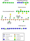
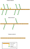
Structure of hemicelluloses found in the cell wall of land plants (Scheller and Ulvskov, 2010). Xyloglucans are represented in Figure 3. Mannan-type hemicellulose can be found as mannans, galactomannans and galactoglucomannans. Xylan-type hemicellulose has been described as xylans, arabinoxylans (AX) and as represented in the figure: glucuronoarabinoxylans (GAXs). Arabinoglucans were recently described in the moss P. patens (Roberts et al., 2018). Structures under braces are only found in monocots. β-(1-4)(1-3)-mixed linkage glucans, not discussed in this review, are not represented. Monosaccharides are represented according to the Symbol Nomenclature for Glycans (SNFG) (Varki et al., 2015), at the exception of the ferulic acid which is not present in the SNFG. Ferulic acid is represented with a black triangle.
Figure 3
Figure 3
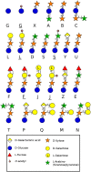
Side chain diversity found in XyG throughout land plant lineages according to Schultink et al. (2014) using the one letter code proposed by Fry et al. (1993). Underlined letters correspond to O-acetylated side chains. Monosaccharides are represented according to the Symbol Nomenclature for Glycans (SNFG) (Varki et al., 2015).
Figure 4
Figure 4
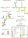
Structure of the different pectin domains found in the cell wall throughout land plant lineages (Wolf et al., 2012). The structural changes of RG-II side chains is presented according to Matsunaga et al. (2004), O'Neill et al. (2004), Pabst et al. (2013), and Ndeh et al. (2017). Linear arabinans were described in N. alata pollen tubes (Lampugnani et al., 2016). Symbols in braces represent methyl decorations: “a,” 0, 1, or 2 methylester(s) that can be found in Lemnoidae, and “b,” methylesters found only in bryophytes (Matsunaga et al., ; Avci et al., 2018). Dha: 3-Deoxy-D-lyxo-hept-2-ulopyranosaric acid, Kdo: 3-Deoxy-D-manno-oct-2-ulopyranosonic acid. Monosaccharides are represented according to the Symbol Nomenclature for Glycans (SNFG) (Varki et al., 2015).
Figure 5
Figure 5
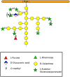
Type-II arabinogalactan structure of a classical AGP according to Nguema-Ona et al. (2012) and Showalter and Basu (2016). Methylesters in braces have been described in mosses and ferns (Fu et al., ; Moller et al., ; Bartels and Classen, 2017). A GlyceroPhosphatidyl Inositol (GPI) anchor can be found in the N-terminal region of the protein but is not represented here. Monosaccharides are represented according to the Symbol Nomenclature for Glycans (SNFG) (Varki et al., 2015).
Figure 6
Figure 6
Structures of extensins described by Carpita et al. (2015) and Showalter and Basu (2016) showing different levels of O-glycosylation. Monosaccharides are represented according to the SNFG (Varki et al., 2015).
Figure 7
Figure 7
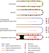
Model presenting the common and the different cell wall polymers found in gametophyte tip-growing cells. Protonemata and rhizoids illustrate the first slow tip-growing cells in P. patens with one cell wall layer composed of weakly methylesterified HGs, mannans, non-fucosylated and GalA containing XyGs and very low level of callose. Evolution has brought more specialized structure devoted to reproduction: pollen tubes. They display striking differences in growth speed: from slow-growing in some gymnosperms, moderate growth in other gymnosperms to fast-growing pollen tubes in angiosperms. In gymnosperms, only one cell wall layer and no callose have been observed. Due to the lack of information, a lot of question marks are shown. The cell wall contains callose, cellulose, and highly methylesterified HGs that is found in the tip and way behind it. In angiosperm monocot non-commelinid and eudicot pollen tubes, two cell wall layers are observed in the shank of the tube and a clear de-methylesterification of HGs is observed in the sub-apical dome. Callose plugs represented in black are regularly synthesized for maintaining the cell in the tip region of the pollen tube. The diameter differences are not in scale but depict variation between those structures. It was estimated based on published pictures. Cellulose is not represented.

Similar articles

Cited by

References

    1. Abel W. O., Knebel W., Koop H. U., Marienfeld J. R., Quader H., Reski R., Schnepf E., Sprlein B. (1989). A cytokinin-sensitive mutant of the moss, Physcomitrella patens, defective in chloroplast division. Protoplasma 152, 1–13. 10.1007/BF01354234 - DOI
    1. Abercrombie J. M., O'Meara B. C., Moffatt A. R., Williams J. H. (2011). Developmental evolution of flowering plant pollen tube cell walls: callose synthase (CalS) gene expression patterns. EvoDevo 2:14. 10.1186/2041-9139-2-14 - DOI - PMC - PubMed
    1. Aloisi I., Cai G., Faleri C., Navazio L., Serafini-Fracassini D., Del Duca S. (2017). Spermine regulates pollen tube growth by modulating Ca2+-dependent actin organization and cell wall structure. Front. Plant Sci. 8:1701. 10.3389/fpls.2017.01701 - DOI - PMC - PubMed
    1. Amborella Genome Project (2013). The Amborella genome and the evolution of flowering plants. Science 342:1241089 10.1126/science.1241089 - DOI - PubMed
    1. Anderson J. R., Barnes W. S., Bedinger P. (2002). 2,6-Dichlorobenzonitrile, a cellulose biosynthesis inhibitor, affects morphology and structural integrity of petunia and lily pollen tubes. J. Plant Physiol. 159, 61–67. 10.1078/0176-1617-00651 - DOI