Review
. 2017 Jan 2:3:16066.
doi: 10.1038/micronano.2016.66. eCollection 2017.

State-of-the-art MEMS and microsystem tools for brain research

Affiliations
Review

State-of-the-art MEMS and microsystem tools for brain research

John P Seymour et al. Microsyst Nanoeng. .

Abstract

Mapping brain activity has received growing worldwide interest because it is expected to improve disease treatment and allow for the development of important neuromorphic computational methods. MEMS and microsystems are expected to continue to offer new and exciting solutions to meet the need for high-density, high-fidelity neural interfaces. Herein, the state-of-the-art in recording and stimulation tools for brain research is reviewed, and some of the most significant technology trends shaping the field of neurotechnology are discussed.

Keywords: MEMS; brain research; electrophysiology; microelectrodes; neural engineering; neuroscience; optoelectrodes; optogenetics.


Conflict of interest statement

FW is the VP of Product Development of Diagnostic Biochips, Inc., a for-profit manufacturer of neurotechnology. The remaining authors declare no conflicts of interest.

Figures

Figure 1
Figure 1
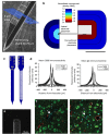
Recording and stimulating technologies vary across scale and degrees of invasiveness. (a) Illustration of the rodent brain and a variety of technologies from electroencephalogram (EEG) to intracortical microelectrodes. (b) High-density systems will increasingly require built-in active electronics to serialize large data streams and reduce the size of the connectors. Sample electrical signals show the amplitudes of various signal sources. The intracortical arrays are often microelectrodes but may also include chemical and optical sensors. (c) Polyimide electrocorticogram (ECoG) for large area mapping. (d) A “Utah array” with 400 μm shank spacing and 100 channels has been used in human studies. (e) Close-packed recording sites with 9×9 μm area and a pitch of 11 μm. (f) MicroLED optoelectrode made from GaN on silicon. (g) Parylene ECoG with greatly improved resolution over EEG and even single-cell capabilities. (h) CMOS integration on probe shaft and backend. (i) Fluidic probe for drug delivery. (j) Active 3D silicon recording system with flexible parylene interconnect.
Figure 2
Figure 2
Seminal work supporting the hypothesis that the tissue response is a function of local device structure. (a and b) Tissue around the end of a thin polymer structure showed significant reduction in encapsulating cells (modified from Ref. 98). (c and d) Tissue response around solid and fine lattice structures showed significant reduction in reactive markers such as CD68 and IgG,. (e and f) Carbon fiber microthreads with an 8-μm diameter reduced tissue reactivity and improved neuron density of microthread. IgG, immunoglobulin G.
Figure 3
Figure 3
Log scale of elastic modulus for many substrates used in implantable arrays. Seminal research has covered inorganic substrates such as silicon, titanium, diamond, zinc oxide, and silicon carbide. Studies on organic substrates have covered carbon fiber, parylene, SU-8,, polyimide,, and silicone.
Figure 4
Figure 4
Example optoelectrodes with integrated waveguides: (ac) Laser diode coupled waveguide probe demonstrating diode directly mounted on neural probe; (d) a digital micromirror directing multi-color light into waveguides terminated with metal-coated corner mirrors; (e) single waveguide on a silicon recording array; (f and g) schematic of multi-color laser diodes coupled from a PCB using graded-index lenses and mixed on the neural probe and micrograph of an actual device; and (h) a 4x4 ZnO array demonstrating a very similar form factor as the Utah array with the added capability of optical stimulation through the ZnO tine and ITO-coated tip.
Figure 5
Figure 5
Fiberless optical stimulation using μLEDs. (a) GaN μLEDs grown on sapphire wafers and transferred onto a polymer substrate by laser-liftoff achieved 50×50 μm2 μLEDs. (b) First demonstration of monolithic integration of multiple GaN μLEDs on silicon neural probes and capable of a 50 μm pitch. Scale=15 μm. (c) In vivo demonstration of same optoelectrode controlling pyramidal cells (PYR) in distinct parts of the CA1 pyramidal cell layer.
Figure 6
Figure 6
Scalability of leading high-density recording systems having integrated digital output. (a) Channel count versus density for three different architectures. Color indicates the input-referred noise (μVrms). Actual channel count was significantly lower compared with the number of available recording sites for the switch-matrix architecture (□). The arrow indicates the direction and color of advancing microsystems. (b) Legend for inset A showing three common architectures discussed in recent work. Table 1 draws further comparison.

References

    1. Allen PG, Collins FS. Toward the final frontier: The human brain. Wall Street Journal 2013.
    1. Azevedo FAC, Carvalho LRB, Grinberg LT et al. Equal numbers of neuronal and nonneuronal cells make the human brain an isometrically scaled up primate brain. Journal of Comparitive Neurology 2009; 513: 532–541. - PubMed
    1. Buzsáki G. Neural syntax: Cell assemblies, synapsembles, and readers. Neuron 2010; 68: 362–385. - PMC - PubMed
    1. Roux L, Stark E, Sjulson L et al. In vivo optogenetic identification and manipulation of GABAergic interneuron subtypes. Current Opinions in Neurobiology 2014; 26C: 88–95. - PMC - PubMed
    1. Fishell G, Heintz N. The neuron identity problem: Form meets function. Neuron 2013; 80: 602–612. - PubMed