Huge Primary Parasitic Leiomyoma in a Postmenopausal Lady: A Rare Presentation
- PMID: 31057977
- PMCID: PMC6463606
- DOI: 10.1155/2019/7683873
Huge Primary Parasitic Leiomyoma in a Postmenopausal Lady: A Rare Presentation
Abstract
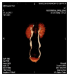
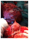
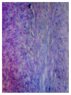
Although uterine myomas are the most common benign tumours of the female pelvis in the reproductive age group, they rarely grow in menopausal women. Parasitic fibroids without prior history of laparoscopic myomectomy are even a rarer presentation particularly in menopausal women. The case presented is a 58-year-old grand-multiparous, menopausal lady with progressive abdominal swelling of three-year duration. She had excision of a huge parasitic fibroid attached to omentum. She had partial omentectomy, total abdominal hysterectomy, and bilateral salpingo-oophorectomy. The parasitic fibroid mass weighed 5.2kg and histopathology confirmed leiomyoma uteri with cystic degeneration and lymph nodes with reactive lymphoid hyperplasia. She had uneventful postoperative recovery and follow-up has so far been uneventful.
Figures


References
-
- Elagwany A. S., Rady H. A., Abdeldayem T. M. A case of parasitic leiomyoma with serpentine omental blood vessels: an unusual variant of uterine leiomyoma. Journal of Taibah University Medical Sciences. 2014;9(4):338–340. doi: 10.1016/j.jtumed.2014.05.002. - DOI
-
- Sukainah S., Nasir T. K., Zulkifli K., et al. Parasitic fibroid: case report and novel approach in reducing incidence of future cases. International Journal of Reproduction, Contraception, Obstetrics and Gynecology. 2016;5:2836–2839. doi: 10.18203/2320-1770.ijrcog20162676. - DOI
Publication types
LinkOut - more resources
Full Text Sources

