Review
. 2019 Apr 18:6:71.
doi: 10.3389/fmed.2019.00071. eCollection 2019.

18F-FDG-PET in Mouse Models of Alzheimer's Disease

Affiliations
Review

18F-FDG-PET in Mouse Models of Alzheimer's Disease

Caroline Bouter et al. Front Med (Lausanne). .

Abstract

Suitable animal models and in vivo biomarkers are essential for development and evaluation of new therapeutic strategies in Alzheimer's disease (AD). 18F-Fluorodeoxyglucose (18F-FDG)-positron-emission tomography (PET) is an imaging biomarker that allows the assessment of cerebral glucose metabolism in vivo. While 18F-FDG-PET/CT is an established tool in the evaluation of AD patients, its role in preclinical studies with AD mouse models remains unclear. Here, we want to review available studies on 18F-FDG-PET/CT in AD mouse models in order to evaluate the method and its impact in preclinical AD research. Only a limited number of studies using 18F-FDG-PET in AD mice were carried out so far showing contradictory findings in cerebral FDG uptake. Methodological differences as well as underlying pathological features of used mouse models seem to be accountable for those varying results. However, 18F-FDG-PET can be a valuable tool in longitudinal in vivo therapy monitoring with a lot of potential for future studies.

Keywords: 18F-FDG-PET; APP; Alzheimer's disease; PET; mouse model; presenilin.


Figures
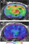
Figure 1
Figure 1
18F-FDG-PET in Tg4-42 mice. Mice were fasted overnight and 18F-FDG (mean 16.2 MBq) was administered intravenously. Scans were performed after an awake uptake period of 45 min. Mice were anesthetized with isoflurane for the injection and during the scans. PET images were acquired on a small animal 1 Tesla nano scan PET/MRI (Mediso, Hungary) for 20 min. A 136 × 131 × 315 matrix with a voxel size of 0.23 × 0.3 × 0.3 mm3 was used. MRI images were used for attenuation correction (matrix 144 × 144 × 163 with a voxel size of 0.5 × 0.5 × 0.6 mm3, TR: 15 ms, TE 2.032 ms and a flip angle of 25°). Fused PET/MRI images are shown in coronal view. (A) 18F-FDG-PET/MRI of a wildtype mouse with homogenous FDG-distribution in all brain areas. (B) 18F-FDG-PET/MRI of an aged Tg4-42 mouse with distinct lower uptake compared to wildtype mice. A, amygdala; C, cortex; H, hypothalamus; Hc, hippocampus; T, thalamus.

References

    1. Kadekaro M, Crane AM, Sokoloff L. Differential effects of electrical stimulation of sciatic nerve on metabolic activity in spinal cord and dorsal root ganglion in the rat. Proc Natl Acad Sci USA. (1985) 82:6010–3. 10.1073/pnas.82.17.6010 - DOI - PMC - PubMed
    1. Sokoloff L. The physiological and biochemical bases of functional brain imaging. Cogn Neurodyn. (2008) 2:1–5. 10.1007/s11571-007-9033-x - DOI - PMC - PubMed
    1. Bloudek LM, Spackman DE, Blankenburg M, Sullivan SD. Review and meta-analysis of biomarkers and diagnostic imaging in Alzheimer's disease. J Alzheimers Dis. (2011) 26:627–45. 10.3233/JAD-2011-110458 - DOI - PubMed
    1. Irizarry MC, Mcnamara M, Fedorchak K, Hsiao K, Hyman BT. APPSw transgenic mice develop age-related A beta deposits and neuropil abnormalities, but no neuronal loss in CA1. J Neuropathol Exp Neurol. (1997) 56:965–73. 10.1097/00005072-199709000-00002 - DOI - PubMed
    1. Sasaki A, Shoji M, Harigaya Y, Kawarabayashi T, Ikeda M, Naito M, et al. . Amyloid cored plaques in Tg2576 transgenic mice are characterized by giant plaques, slightly activated microglia, and the lack of paired helical filament-typed, dystrophic neurites. Virchows Arch. (2002) 441:358–67. 10.1007/s00428-002-0643-8 - DOI - PubMed

LinkOut - more resources