Review
. 2019 Apr 1:2019:8973076.
doi: 10.1155/2019/8973076. eCollection 2019.

Transcriptional Regulation of Ovarian Steroidogenic Genes: Recent Findings Obtained from Stem Cell-Derived Steroidogenic Cells

Affiliations
Review

Transcriptional Regulation of Ovarian Steroidogenic Genes: Recent Findings Obtained from Stem Cell-Derived Steroidogenic Cells

Takashi Yazawa et al. Biomed Res Int. .

Abstract

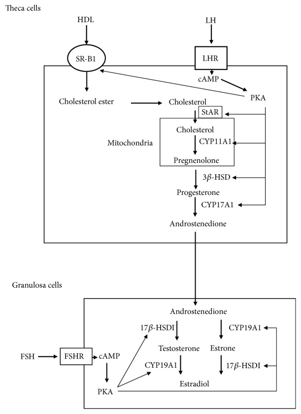
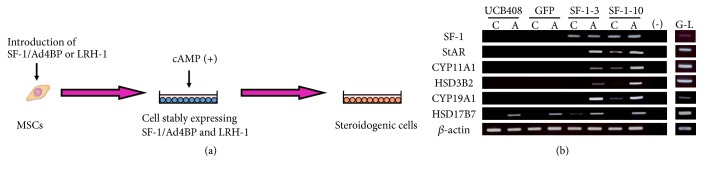
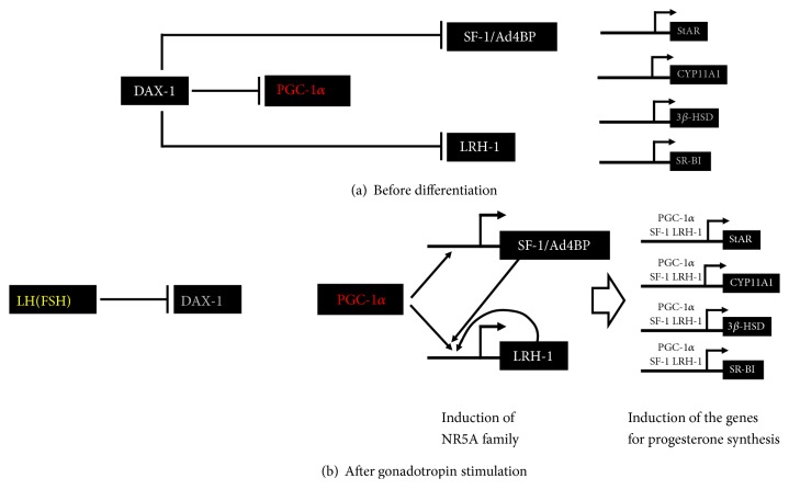
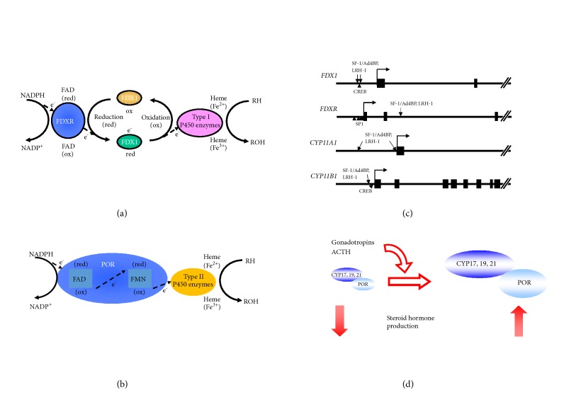
Ovaries represent one of the primary steroidogenic organs, producing estrogen and progesterone under the regulation of gonadotropins during the estrous cycle. Gonadotropins fluctuate the expression of various steroidogenesis-related genes, such as those encoding steroidogenic enzymes, cholesterol deliverer, and electronic transporter. Steroidogenic factor-1 (SF-1)/adrenal 4-binding protein (Ad4BP)/NR5A1 and liver receptor homolog-1 (LRH-1) play important roles in these phenomena via transcriptional regulation. With the aid of cAMP, SF-1/Ad4BP and LRH-1 can induce the differentiation of stem cells into steroidogenic cells. This model is a useful tool for studying the molecular mechanisms of steroidogenesis. In this article, we will provide insight into the transcriptional regulation of steroidogenesis-related genes in ovaries that are revealed from stem cell-derived steroidogenic cells. Using the cells derived from the model, novel SF-1/Ad4BP- and LRH-1-regulated genes were identified by combined DNA microarray and promoter tiling array analyses. The interaction of SF-1/Ad4BP and LRH-1 with transcriptional regulators in the regulation of ovarian steroidogenesis was also revealed.


Figures

Figure 1
Figure 1
Schematic diagram of two-cell–two-gonadotropin theory in ovarian steroidogenesis. Theca cells autonomously produce androstenedione from cholesterol via the positive regulation of steroidogenic enzymes by LH/cAMP/PKA pathway. Granulosa cells convert androstenedione into estradiol. It is promoted by FSH/cAMP/PKA pathway.
Figure 2
Figure 2
Differentiation of MSCs into steroidogenic cells. (a) Schematic diagram of induction of steroidogenic cells from MSCs by introduction of SF-1/Ad4BP or LRH, and cAMP-treatment. (b) Differentiation of UCB-MSCs into luteal-like cells by SF-1/Ad4BP. RT-PCR analysis of each gene in each cell with or without 8-bromo-cAMP for 2 d. Lanes G–L are granulosa-luteal cells from women undergoing oocyte retrieval for in vitro fertilization.
Figure 3
Figure 3
Regulation of granulosa cell differentiation by DAX-1 and PGC-1α via SF-1/Ad4BP and LRH-1. (a) During follicular development, the activities of SF-1/Ad4BP, LRH-1, and PGC-1α are repressed by DAX-1 in undifferentiated granulosa cells. Therefore, transcription of steroidogenic genes hardly occurred. (b) Gonadotropin induces the differentiation of granulosa cells into progesterone-producing cells. At this time, PGC-1α is released from DAX-1-mediated suppression via its reduction. This PGC-1α activation induces SF-1/Ad4BP and LRH-1. In particular, LRH-1 is highly induced by positive-feedback loop. Then, NR5A family proteins couple with PGC-1α to induce steroidogenic genes, such as StAR, CYP11A1, 3β-HSD, and SR-BI.
Figure 4
Figure 4
P450 enzymes and their redox partners in steroidogenesis. (a) Schematic diagram of electron transfer from NADPH to mitochondrial type I P450 enzymes by FDXR and FDX1. At first, NADPH passes the electrons to the nonreduced (ox) FAD moiety of FDXR. Then, reduced (red) FDXR passes them to FDX1, followed by the transfer to P450 enzymes and hydroxylation of cholesterol and its metabolites. (b) Schematic diagram of electron transfer from NADH to microsomal type II P450 enzymes by POR. POR receives the electrons from NADPH by using its FAD moiety, and red FAD passes them to its FMN. Then, electrons are transferred to P450 enzymes to activate the steroid hormone production. (c) The binding sites of NR5A family and other transcription factors, which are essential for the transcription of FDX, FDXR, and ovarian type I P450 genes. (d) Pituitary hormones promote the steroidogenesis by enhancing the transcription of both mitochondrial P450 enzymes and POR.

References

    1. Miller W. L., Auchus R. J. The molecular biology, biochemistry, and physiology of human steroidogenesis and its disorders. Endocrine Reviews. 2011;32(1):81–151. doi: 10.1210/er.2010-0013. - DOI - PMC - PubMed
    1. Miller W. L. Steroidogenesis: Unanswered Questions. Trends in Endocrinology & Metabolism. 2017;28(11):771–793. doi: 10.1016/j.tem.2017.09.002. - DOI - PubMed
    1. Connelly M. A., Williams D. L. SR-BI and cholesterol uptake into steroidogenic cells. Trends in Endocrinology & Metabolism. 2003;14(10):467–472. doi: 10.1016/j.tem.2003.10.002. - DOI - PubMed
    1. Connelly M. A. SR-BI-mediated HDL cholesteryl ester delivery in the adrenal gland. Molecular and Cellular Endocrinology. 2009;300(1-2):83–88. doi: 10.1016/j.mce.2008.09.011. - DOI - PubMed
    1. Stocco D. M., Zhao A. H., Tu L. N., Morohaku K., Selvaraj V. A brief history of the search for the protein(s) involved in the acute regulation of steroidogenesis. Molecular and Cellular Endocrinology. 2017;441:7–16. doi: 10.1016/j.mce.2016.07.036. - DOI - PMC - PubMed

LinkOut - more resources