Comparative Study
. 2019 Apr 19:(146):10.3791/59290.
doi: 10.3791/59290.

Rapid Lipid Droplet Isolation Protocol Using a Well-established Organelle Isolation Kit

Affiliations
Comparative Study

Rapid Lipid Droplet Isolation Protocol Using a Well-established Organelle Isolation Kit

Jascha Brettschneider et al. J Vis Exp. .

Abstract

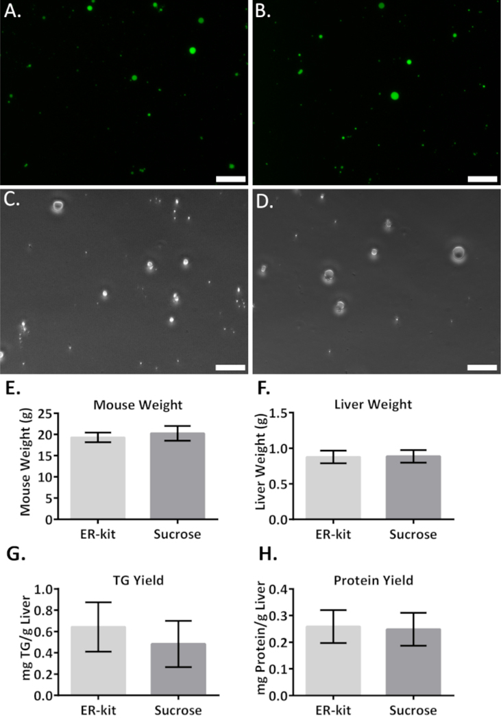
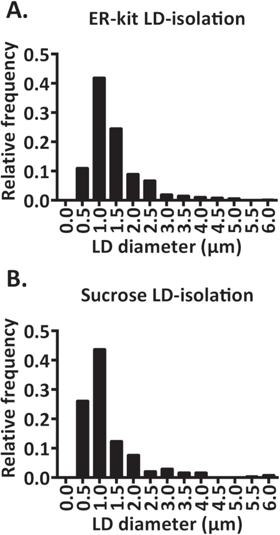
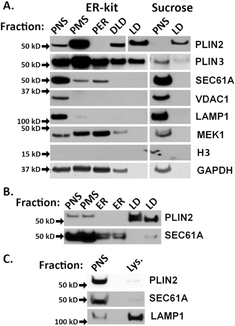
Lipid droplets (LDs) are bioactive organelles found within the cytosol of the most eukaryotic and some prokaryotic cells. LDs are composed of neutral lipids encased by a monolayer of phospholipids and proteins. Hepatic LD lipids, such as ceramides, and proteins are implicated in several diseases that cause hepatic steatosis. Although previous methods have been established for LD isolation, they require a time-consuming preparation of reagents and are not designed for the isolation of multiple subcellular compartments. We sought to establish a new protocol to enable the isolation of LDs, endoplasmic reticulum (ER), and lysosomes from a single mouse liver. Further, all reagents used in the protocol presented here are commercially available and require minimal reagent preparation without sacrificing LD purity. Here we present data comparing this new protocol to a standard sucrose gradient protocol, demonstrating comparable purity, morphology, and yield. Additionally, we can isolate ER and lysosomes using the same sample, providing detailed insight into the formation and intracellular flux of lipids and their associated proteins.


Figures

Figure 1:
Figure 1:. Reference pictures taken during the LD isolation using an ER isolation kit.
(A) Diced 5 mm liver pieces in a 5 cm Petri dish. (B) Liver homogenate after homogenization in a Dounce homogenizer. (C) Liver homogenate after centrifugation at 1,000 × g; note the slight lipid layer on top, the PNS, and the pellet containing cell debris and nuclear fraction. (D) LD layer after ultracentrifugation at 100,000 × g; note the prominent lipid layer on top, the PER, and the ER fraction on the bottom. (E) Washed LD fraction before the final removal of the PBS.
Figure 2:
Figure 2:. Micrograph and quantitative analysis comparing the LD fraction isolated with an ER kit and the LD fraction isolated with sucrose.
Representative 20x fluorescent micrograph of (A) a DPBDI-stained LD fraction isolated with an ER kit and (B) LDs isolated with sucrose. (C) Brightfield micrograph of the LD fraction isolated with an ER kit and (D) the LD fraction isolated with sucrose. The scale bar = 20 μm. (E) Mouse weight and (F) liver weight. (G) TG yield from purified LDs normalized to liver weight. (H) Protein yield from purified LDs normalized to liver weight. The data are mean ± SEM (N = 4).
Figure 3:
Figure 3:. Frequency distribution of LD sizes.
Samples from four different mice were stained with DPBDI and representative 20x fields were quantified. The diameters were measured using an image analysis software. (A) Frequency (fraction) distribution of an LD fraction isolated using an ER-kit. (B) Frequency (fraction) distribution of an LD fraction isolated using sucrose gradient density.
Figure 4:
Figure 4:. Analysis of LD purity by immunoblotting.
(A) To compare the adapted protocol of the ER kit LD isolation and the sucrose isolation protocol, 20 μg of protein per sample was immunoblotted. The following samples were loaded: postnuclear supernatant (PNS), postmitochondrial supernatant (PMS), postendoplasmic reticulum supernatant (PER), crude LDs (CLD), and LDs (LD). Samples were immunoblotted for LDs (PLIN2 and PLIN3), ER (SEC61A), mitochondria (VDAC1), lysosome (LAMP1), cell membrane (MEK1), nuclei (histone H3) and cytosol (GAPDH) markers. (B) Purity analysis of an ER fraction after further purification of the ER using the ER kit LD isolation protocol (see the Table of Materials). PNS, PMS, ER, and LDs were loaded and immunoblotted for LDs (PLIN2) and ER (SEC61A). (C) Purity analysis of a lysosomal fraction after further purification of lysosomes using the lysosome enrichment kit protocol (see the Table of Materials). PNS and a lysosomal fraction were loaded and immunoblotted for LDs (PLIN2), ER (SEC61A), and lysosomes (LAMP1).

References

    1. Brasaemle DL, Wolins NE Isolation of Lipid Droplets from Cells by Density Gradient Centrifugation. Current Protocols in Cell Biology. 72, 3.15.11–13.15.13 (2016). - PMC - PubMed
    1. Ding Y et al. Isolating lipid droplets from multiple species. Nature Protocols. 8 (1), 43–51 (2013). - PubMed
    1. Gao Q, Goodman JM The lipid droplet-a well-connected organelle. Frontiers in Cell and Developmental Biology. 3, 49 (2015). - PMC - PubMed
    1. Correnti JM et al. Ethanol and C2 ceramide activate fatty acid oxidation in human hepatoma cells. Scientific Reports. 8 (1), 12923 (2018). - PMC - PubMed
    1. Williams B et al. A novel role for ceramide synthase 6 in mouse and human alcoholic steatosis. FASEB Journal. 32 (1), 130–142 (2018). - PMC - PubMed

Publication types