. 2019 Jun;19(6):4897-4905.
doi: 10.3892/mmr.2019.10158. Epub 2019 Apr 12.

Endophilin 1 knockdown prevents synaptic dysfunction induced by oligomeric amyloid β

Affiliations

Endophilin 1 knockdown prevents synaptic dysfunction induced by oligomeric amyloid β

Yichen Yin et al. Mol Med Rep. 2019 Jun.

Abstract

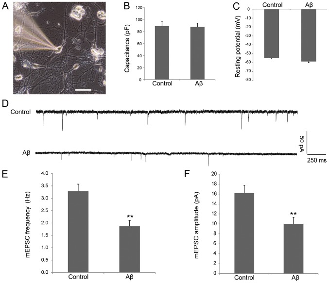
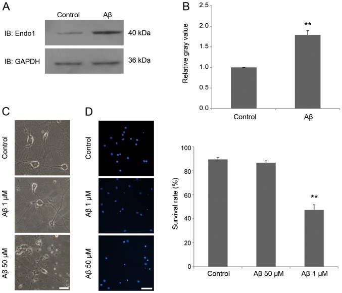
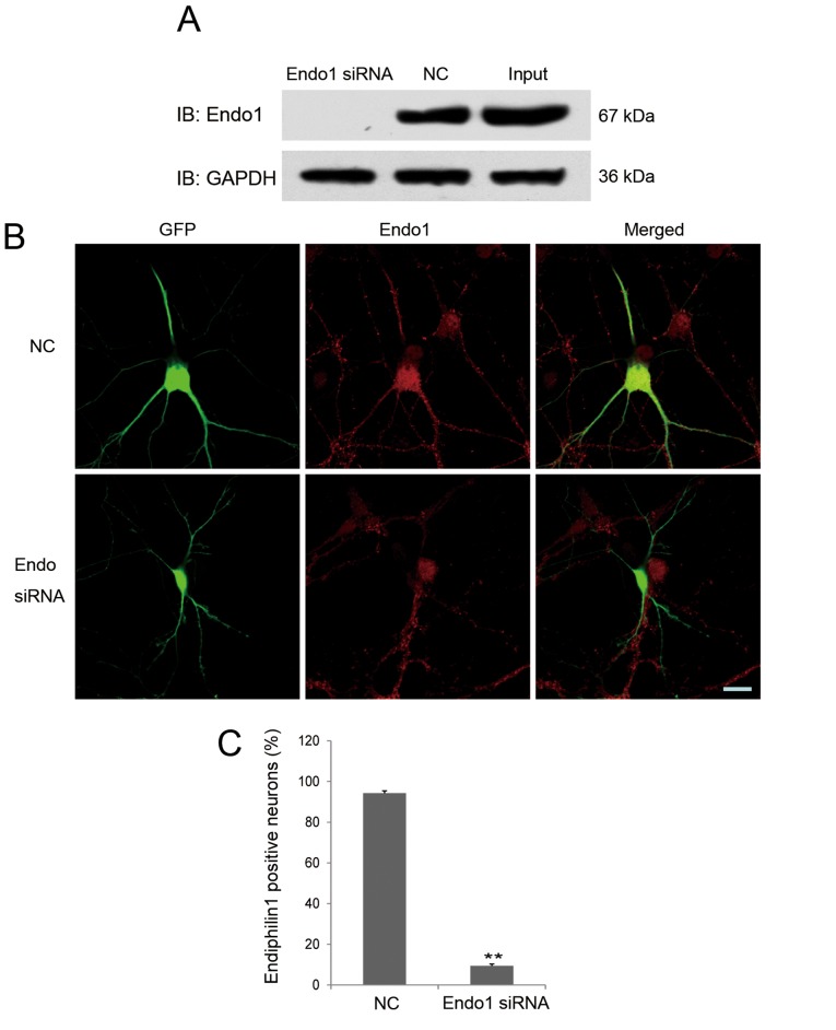
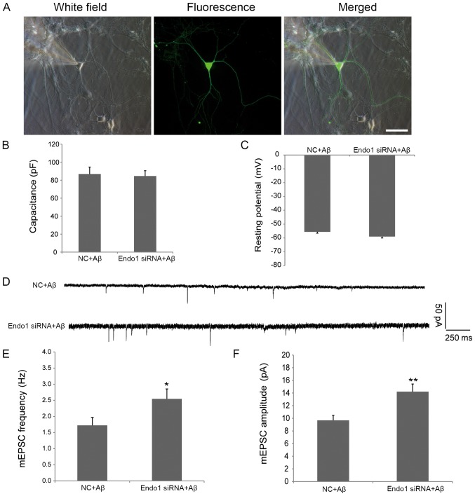
Amyloid β (Aβ) has been reported to have an important role in the cognitive deficits of Alzheimer's disease (AD), as oligomeric Aβ promotes synaptic dysfunction and triggers neuronal death. Recent evidence has associated an endocytosis protein, endophilin 1, with AD, as endophilin 1 levels have been reported to be markedly increased in the AD brain. The increase in endophilin 1 levels in neurons is associated with an increase in the activation of the stress kinase JNK, with subsequent neuronal death. In the present study, whole‑cell patch‑clamp recording demonstrated that oligomeric Aβ caused synaptic dysfunction and western blotting revealed that endophilin 1 was highly expressed prior to neuronal death of cultured hippocampal neurons. Furthermore, RNA interference and electrophysiological recording techniques in cultured hippocampal neurons demonstrated that knockdown of endophilin 1 prevented synaptic dysfunction induced by Aβ. Thus, a potential role for endophilin 1 in Aβ‑induced postsynaptic dysfunction has been identified, indicating a possible direction for the prevention of postsynaptic dysfunction in cognitive impairment and suggesting that endophilin may be a potential target for the clinical treatment of AD.


Figures

Figure 1.
Figure 1.
Electrophysiology of cultured hippocampal neurons treated with oligomeric 1 µM Aβ. (A) Image of a neuron obtained from patch recording captured using an inverted fluorescence microscope. Scale bar, 50 µm. Bar plots represent the mean values of resting (B) capacitance and (C) membrane potentials of patched neurons in the control and oligomeric Aβ-processed groups. n=10 cells per group. (D) mEPSC tracings are shown for control and oligomeric Aβ-treated neurons. (E) mEPSC frequency in control and oligomeric Aβ-treated neurons. (F) mEPSC amplitude in control and oligomeric Aβ-treated neurons. n=16 cells per group, **P<0.01 vs. control. Aβ, amyloid β; mEPSC, miniature excitatory postsynaptic current.
Figure 2.
Figure 2.
Increased expression of endophilin 1 in neuronal synaptic dysfunction induced by oligomeric Aβ. (A) Hippocampal neurons were treated with Aβ. After 48 h, endophilin 1 and GAPDH were probed using the designated antibodies. (B) Relative expression of endophilin 1 in the two groups n=4. (C) Photomicrographs illustrating hippocampal neurons treated with 0, 1 and 50 µM oligomeric Aβ. Scale bar, 25 µm. (D) DAPI staining assay showing survival of hippocampal neurons treated with 0, 1 and 50 µM oligomeric Aβ. Scale bar, 50 µm. n=5 per group. **P<0.01 vs. control. Aβ, amyloid β; IB, immunoblot; Endo 1, endophilin 1.
Figure 3.
Figure 3.
Verification of the efficacy of endophilin 1 siRNA. (A) 293 cells were co-transfected with endophilin 1-pEGFP-C1 plasmids and Endo1 siRNA or NC. After 48 h, endophilin 1 and GAPDH as a control in the cell lysates were probed by the designated antibodies. (B) Images of neurons co-transfected with green fluorescent protein and NC or Endo1 siRNA. Cultures were fixed after transfection for 2–3 days and stained using endophilin 1 antibody (red). Scale bar, 20 µm. (C) Percentage of endophilin 1-positive neurons in the two groups. n=4, **P<0.01. Endo1, endophilin 1; siRNA, small interfering RNA; NC, negative control; IB, immunoblot; GFP, green fluorescent protein; Input, endophilin 1-pEGFP-C1 plasmid transfection only.
Figure 4.
Figure 4.
Electrophysiology of endophilin 1 knockdown neurons processed by oligomeric Aβ. (A) Image of a neuron obtained from patch recording. Scale bar, 50 µm. Mean values of (B) capacitance and (C) resting membrane potentials of patched neurons in NC + Aβ and Endo1 siRNA + Aβ groups. n=10 cells per group. (D) mEPSC tracings from NC + Aβ and Endo1 siRNA + Aβ groups. (E) mEPSC frequency and (F) amplitude in NC + Aβ and Endo1 siRNA + Aβ group, n=16 cells per group, *P<0.05, **P<0.01 vs. NC + Aβ. NC, negative control; Aβ, amyloid β; Endo1, endophilin 1; siRNA, small interfering RNA; mEPSC, miniature excitatory postsynaptic current.

References

    1. delEtoile J, Adeli H. Graph theory and brain connectivity in Alzheimer's disease. Neuroscientist. 2017;23:616–626. doi: 10.1177/1073858417702621. - DOI - PubMed
    1. Lane CA, Hardy J, Schott JM. Alzheimer's disease. Eur J Neurol. 2018;25:59–70. doi: 10.1111/ene.13439. - DOI - PubMed
    1. Luo J, Wärmländer SK, Graslund A, Abrahams JP. Cross-interactions between the alzheimer disease amyloid-β peptide and other amyloid proteins: A further aspect of the amyloid cascade hypothesis. J Biol Chem. 2016;291:16485–16493. doi: 10.1074/jbc.R116.714576. - DOI - PMC - PubMed
    1. Jamasbi E, Wade JD, Separovic F, Hossain MA. Amyloid Beta (Aβ) Peptide and factors that play important roles in Alzheimer's disease. Curr Med Chem. 2016;23:884–892. doi: 10.2174/0929867323666160229113911. - DOI - PubMed
    1. Pini L, Pievani M, Bocchetta M, Altomare D, Bosco P, Cavedo E, Galluzzi S, Marizzoni M, Frisoni GB. Brain atrophy in Alzheimer's disease and aging. Ageing Res Rev. 2016;30:25–48. doi: 10.1016/j.arr.2016.01.002. - DOI - PubMed

MeSH terms