. 2019 Jun;19(6):5424-5432.
doi: 10.3892/mmr.2019.10208. Epub 2019 Apr 30.

Ghrelin regulates sepsis‑induced rat acute gastric injury

Affiliations

Ghrelin regulates sepsis‑induced rat acute gastric injury

Bin Li et al. Mol Med Rep. 2019 Jun.

Abstract

Ghrelin, a peptide expressed in the gastric mucosa, has an essential role in sustaining the normal function of the digestive system. Sepsis is one of the primary causes of mortality in intensive care units and can lead to multiple organ dysfunction, especially in the gastrointestinal system. The aim of the present study was to explore the effect of ghrelin on gastric blood flow in a rat model of sepsis, as well as the effect of ghrelin on the expression of the apoptotic markers, B‑cell lymphoma 2 (Bcl‑2) and Bcl‑2‑associated X (Bax), in gastric tissues. The sepsis model was established using cecal ligation and puncture (CLP). The expression levels of apoptosis‑related factors in gastric epithelial cell were determined by immunohistochemistry, reverse transcription quantitative‑PCR and western blotting. Collectively, the present results suggested that ghrelin administration attenuated sepsis symptoms induced by CLP. Blood flow in the stomach greater curvature was significantly higher in the CLP‑induced sepsis group rats (284.3±95.7 perfusion units) compared with the sham operation group (317.8±5.2 perfusion units; P<0.05), whereas there was no difference between the CLP group treated with ghrelin (377.8±99.0 perfusion units) and the sham rats. Ghrelin administration also reduced the secretion of pro‑inflammatory cytokines compared with the CLP‑induced sepsis group rats. In addition, CLP significantly reduced the expression of Bcl‑2 and enhanced the expression of the pro‑apoptotic proteins, Bax and cleaved caspase‑3; whereas, ghrelin application reversed the effects of CLP on these apoptosis‑associated proteins. In conclusion, the present study revealed that ghrelin has the ability to increase blood flow in the gastrointestinal tract in a sepsis model and can also regulate the expressions of apoptosis‑associated factors in gastric tissues. These results suggest that ghrelin could be a novel treatment for sepsis‑induced gastric injury.


Figures

Figure 1.
Figure 1.
Ghrelin levels in the septic rat. (A) mRNA levels of ghrelin detected in the isolated gastric epithelial cells from the indicated group of the rats. A representative semi-quantitative polymerase chain reaction result is presented; 3 rats in each group were used to detect the mRNA levels in sham and CLP-treated rats. Data were normalized to GAPDH and expressed as the mean ± standard error. (B) Ghrelin levels detected in stomach mucosa. (C) Ghrelin levels in the plasma. Each data point represents a rat. *P<0.05, as indicated. CLP, cecal ligation and puncture.
Figure 2.
Figure 2.
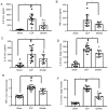
Tail blood pressure (mean) and blood flow in the stomach. (A) Blood pressure of rats in each group. At 2 h post-CLP, the tail blood pressure was measured in the rats of each group. (B) Blood perfusion to the stomach greater curvature; blood perfusion (blood flow) was monitored at 20 h post-CLP surgery. All data are expressed as the mean ± standard error. Each data point represents a rat. *P<0.05, as indicated. CLP, cecal ligation and puncture.
Figure 3.
Figure 3.
Pro-inflammatory cytokine levels in serum and tissues from sepsis model rats. Alterations in the levels of IL-1β, TNF-α and IL-6 in the serum and the stomach lysate of sham-operated animals (Sham group), and animals with CLP treated with normal saline (CLP group) or ghrelin (ghrelin group) at 20 h post-surgery. (A) IL-6, (B) TNF-α and (C) IL-1β levels in serum. (D) IL-6, (E) TNF-α and (F) IL-1β levels in tissue from the stomach lysate. Data are presented as the mean ± standard error (n=12/group) and compared by one-way analysis of variance and the Student-Newman-Keuls test. *P<0.05, as indicated. IL, interleukin; TNF-α, tumor necrosis factor-α; CLP, cecal ligation and puncture; conc., concentration.
Figure 4.
Figure 4.
Effect of ghrelin on the expression levels of apoptosis-associated proteins in the sepsis model. (A) Representative immunohistochemistry staining in sham, CLP and ghrelin-treated rats (rows 1 and 2). Immunofluorescent staining for cleaved caspase-3 in the stomach (row 3). Green staining, cleaved caspase 3; blue staining, nuclear staining with DAPI. (B) Western blot analysis of Bcl-2, Bax and cleaved caspase-3 in the sham, CLP and ghrelin-treated rats. β-actin was used as the loading control. The blots are representative of at least 3 independent experiments. CLP, cecal ligation and puncture; Bcl-2, B-cell lymphoma 2; Bax, Bcl-2-associated X.
Figure 5.
Figure 5.
Ghrelin prevents LPS-induced GEC apoptosis. (A) Naïve GECs (2×10⁵) were cultured in 24-well plates and stimulated by LPS with or without ghrelin. Following the 2, 4 and 8 h incubation periods, the (B) percentage of apoptotic GECs was assessed by staining the cells with Annexin V and PI followed by flow cytometric analysis. Data presented as the mean ± standard error (n=3/group). *P<0.05. LPS, lipopolysaccharide; GEC, gastric epithelial cell; PI, propidium iodide; FSC, forward scatter; SSC, side scatter.

References

    1. Lever A, Mackenzie I. Sepsis: Definition, epidemiology, and diagnosis. BMJ. 2007;335:879–83. doi: 10.1136/bmj.39346.495880.AE. - DOI - PMC - PubMed
    1. Martin GS. Sepsis, severe sepsis and septic shock: Changes in incidence, pathogens and outcomes. Expert Rev Anti Infect Ther. 2012;10:701–706. doi: 10.1586/eri.12.50. - DOI - PMC - PubMed
    1. Singer M, Deutschman CS, Seymour CW, Shankar-Hari M, Annane D, Bauer M, Bellomo R, Bernard GR, Chiche JD, Coopersmith CM, et al. The third international consensus definitions for sepsis and septic shock (Sepsis-3) JAMA. 2016;315:801–810. doi: 10.1001/jama.2016.0287. - DOI - PMC - PubMed
    1. Kennelly PJ, Martin-Loeches I. Long term mortality following sepsis. Ann Transl Med. 2016;4:387. doi: 10.21037/atm.2016.08.31. - DOI - PMC - PubMed
    1. Lang CH, Bagby GJ, Ferguson JL, Spitzer J. Cardiac output and redistribution of organ blood flow in hypermetabolic sepsis. Am J Physiol. 1984;246:R331–R337. - PubMed

MeSH terms