. 2019 May 29;141(21):8450-8461.
doi: 10.1021/jacs.8b13506. Epub 2019 May 15.

Ipomoeassin F Binds Sec61α to Inhibit Protein Translocation

Affiliations

Ipomoeassin F Binds Sec61α to Inhibit Protein Translocation

Guanghui Zong et al. J Am Chem Soc. .

Abstract

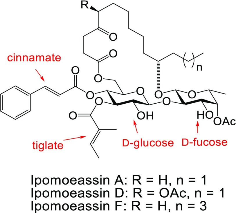
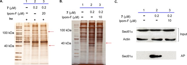
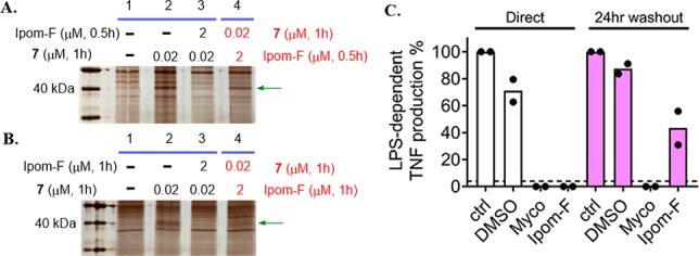
Ipomoeassin F is a potent natural cytotoxin that inhibits growth of many tumor cell lines with single-digit nanomolar potency. However, its biological and pharmacological properties have remained largely unexplored. Building upon our earlier achievements in total synthesis and medicinal chemistry, we used chemical proteomics to identify Sec61α (protein transport protein Sec61 subunit alpha isoform 1), the pore-forming subunit of the Sec61 protein translocon, as a direct binding partner of ipomoeassin F in living cells. The interaction is specific and strong enough to survive lysis conditions, enabling a biotin analogue of ipomoeassin F to pull down Sec61α from live cells, yet it is also reversible, as judged by several experiments including fluorescent streptavidin staining, delayed competition in affinity pulldown, and inhibition of TNF biogenesis after washout. Sec61α forms the central subunit of the ER protein translocation complex, and the binding of ipomoeassin F results in a substantial, yet selective, inhibition of protein translocation in vitro and a broad ranging inhibition of protein secretion in live cells. Lastly, the unique resistance profile demonstrated by specific amino acid single-point mutations in Sec61α provides compelling evidence that Sec61α is the primary molecular target of ipomoeassin F and strongly suggests that the binding of this natural product to Sec61α is distinctive. Therefore, ipomoeassin F represents the first plant-derived, carbohydrate-based member of a novel structural class that offers new opportunities to explore Sec61α function and to further investigate its potential as a therapeutic target for drug discovery.


Conflict of interest statement

The authors declare no competing financial interest.

Figures

Figure 1
Figure 1
Structures of ipomoeassins A, D, and F.
Scheme 1
Scheme 1. Synthesis of Ipomoeassin F Probes 3, 5, and 7
Reagents and conditions: (a) 4-propargyloxycinnamic acid (2), DCC, DMAP, CH2Cl2, 0 °C → RT, overnight, 90%; (b) TBAF, THF, −10 °C, 6 h, 80%; (c) CuSO4, sodium ascorbate, CH2Cl2/t-BuOH, 3:2.
Figure 2
Figure 2
SDS-PAGE images (silver staining) for affinity pulldown using probe 7 with (A) or without (B) photocleavage. Red arrows indicate specifically pulled down protein species. (C) Target validation by Western blot with a Sec61α antibody. Ipom-F, ipomoeassin F; AP, affinity pulldown.
Figure 3
Figure 3
Structures of two inactive derivatives of ipomoeassin F.
Figure 4
Figure 4
Structures of fluorescent derivatives of ipomoeassin F.
Figure 5
Figure 5
Cell imaging studies with fluorescent probe 12 in MDA-MB-231 cells. Rhodamine-conjugated ipomoeassin F analogue 12 (0.2 μM) was added to cells, and after 1 h, cells were imaged to analyze localization of 12 relative to ER staining.
Figure 6
Figure 6
Sheep rough microsomes were preincubated with ipomoeassin F (Ipom-F) or mycolactone (Myco) at 10 μM. Subsequently 100 nM photocotransin CT7 was added and samples were photolyzed. The covalent CT7/Sec61α adduct was detected using click chemistry to install a rhodamine-azide reporter and in-gel fluorescent scanning.
Figure 7
Figure 7
Ipomoeassin F inhibits Sec61-mediated protein translocation into and across the ER in a substrate-selective manner. Phosphorimages of membrane-associated in vitro products resolved by SDS-PAGE together with outline structures of the protein substrates generated in the presence or absence of ipomoeassin F (Ipom-F) or mycolactone (Myco): secretory proteins (A) bovine preprolactin, PPL, and (B) yeast prepro-alpha-factor, ppαF; tail-anchored proteins (C) C-terminally tagged Sec61β subunit, Sec61βOPG2, and (D) C-terminally tagged cytochrome b5, Cytb5OPG2; type I single pass transmembrane protein (TMP) (E) vascular cell adhesion molecule 1, VCAM1; type II single pass TMP (F) isoform 2 (residues 17 to 232) of the short form of HLA class II histocompatibility antigen gamma chain, Ii; type III single pass TMP (G) glycophorin C, GypC; short secretory protein (H) C-terminally tagged Hyalophora cecropia preprocecropin A, ppcecAOPG2. Samples were treated with endoglycosidase H (Endo H) where indicated to distinguish N-glycosylated (XGly) from nonglycosylated (0Gly) products. nc, signal sequence not cleaved; sc, signal sequence cleaved; TM1, transmembrane domain 1. Proteins are of human origin unless otherwise stated.
Figure 8
Figure 8
Structures of ipomoeassin F analogues 1315.
Figure 9
Figure 9
Differential effect of acute ipomoeassin F treatment on protein secretion and cell viability. Bioluminescence outputs from secretion sensor proteins (●) and cellular viability based on total well ATP content (○). (A) secNLuc-ATZ secretion and cell viability measured in U2-OS cells after 24 h. (B) GLuc secretion and cell viability measured in SH-SY5Y cells after 48 h. Error bars represent the SEM for n = 2 or 3 replicates.
Figure 10
Figure 10
HCT-116 cells were washed twice with PBS and incubated in the presence of indicated concentrations of compounds under media lacking methionine and cysteine for 30 min. 35S-labeled methionine and cysteine were then introduced to the media, and cells incubated for a further 90 min. Cells were harvested by scraping into ice cold PBS and analyzed by SDS-PAGE and autoradiography. (A) The collected cells were subsequently homogenized and analyzed by SDS-PAGE and autoradiography. (B) As for (A) but the samples are the cytosolic contents of harvested cells following partial permeabilization with 0.15% digitonin. (C) As for (A) but the samples are from TCA-precipitated culture medium from the same experiment. Ipom-F denotes ipomoeassin F. AprA denotes control samples treated with apratoxin A to block protein translocation into the ER. CHX denotes samples treated with cycloheximide and chloramphenicol to inhibit total cellular protein synthesis. Cotransin CT8 is a substrate-selective inhibitor of protein translocation into the ER.
Figure 11
Figure 11
Compared effects of ipomoeassin F and synthetic mycolactone (A/B form) in assays of CD62L expression (A) and activation-induced production of IL-2 (B) by Jurkat T cells. Data are means ± SEM of biological duplicates and are representative of two independent experiments.
Figure 12
Figure 12
Human HEK293 cells or cells engineered to express Sec61α alleles bearing indicated point mutations were treated with increasing concentrations of ipomoeassin F (A) or mycolactone (B), and cell viability was assayed at 72 h with the Alamar Blue assay (mean ± SD; n = 4 technical quadruplicates). Parental or HCT116 cells containing mutations in Sec61A1 were seeded at 3 × 10⁴/well in 96-well plates in triplicate. Ipomoeassin F (C) or mycolactone (D) were added at various concentrations with the carrier DMSO used as a control. Cell viability was measured at 96 h with the Alamar Blue assay (mean ± SEM of n = 3 independent experiments performed in triplicate).
Figure 13
Figure 13
SDS-PAGE images (silver staining) for affinity pulldown using probe 7 with 0.5 h (A) or 1 h (B) competition in both regular (lane 3) and reverse (lane 4) order. Green arrows indicate the protein band for Sec61α. (C) TNF abundance in RAW264.7 cell supernatants 4 h after stimulation with 100 ng/mL LPS in the presence of medium alone (ctrl), DMSO, 125 ng/mL mycolactone (Myco), or 250 nM ipomoeassin F (Ipom-F). Cells were either preincubated for 1 h prior to addition of LPS (“direct”) or incubated for 1 h, washed, and allowed to recover for 24 h in complete medium before addition of LPS (“24 h washout”). TNF was quantified by ELISA in triplicate, and the dashed line shows the detection limit of the assay. Controls for each condition without LPS stimulation did not reach this detectability threshold. For each replicate, data were normalized to control levels and the mean of n = 2 independent cellular assays is presented.

References

    1. Newman D. J.; Cragg G. M. Natural Products as Sources of New Drugs from 1981 to 2014. J. Nat. Prod. 2016, 79, 629–661. 10.1021/acs.jnatprod.5b01055. - DOI - PubMed
    1. Cragg G. M.; Grothaus P. G.; Newman D. J. Impact of Natural Products on Developing New Anti-Cancer Agents. Chem. Rev. 2009, 109, 3012–3043. 10.1021/cr900019j. - DOI - PubMed
    1. Carlson E. E. Natural Products as Chemical Probes. ACS Chem. Biol. 2010, 5, 639–653. 10.1021/cb100105c. - DOI - PMC - PubMed
    1. Rizzo S.; Waldmann H. Development of a Natural-Product-Derived Chemical Toolbox for Modulation of Protein Function. Chem. Rev. 2014, 114, 4621–4639. 10.1021/cr400442v. - DOI - PubMed
    1. Pye C. R.; Bertin M. J.; Lokey R. S.; Gerwick W. H.; Linington R. G. Retrospective analysis of natural products provides insights for future discovery trends. Proc. Natl. Acad. Sci. U. S. A. 2017, 114, 5601–5606. 10.1073/pnas.1614680114. - DOI - PMC - PubMed

Publication types

MeSH terms