. 2019 May 6;14(5):e0215779.
doi: 10.1371/journal.pone.0215779. eCollection 2019.

Genetic analysis of ATP7B in 102 south Indian families with Wilson disease

Affiliations

Genetic analysis of ATP7B in 102 south Indian families with Wilson disease

Nivedita Singh et al. PLoS One. .

Abstract

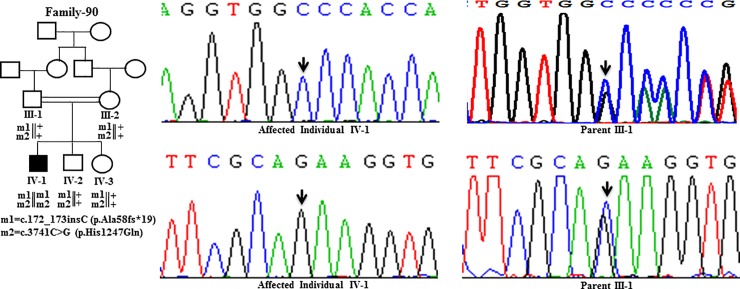
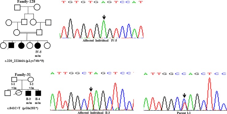
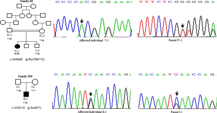
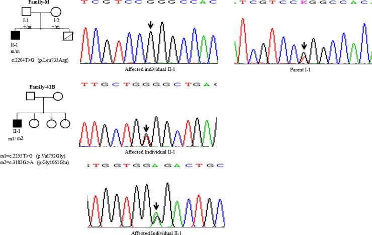
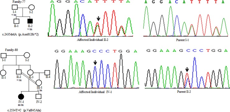
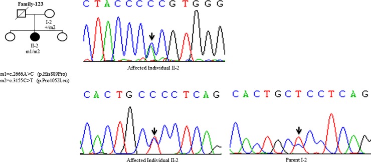
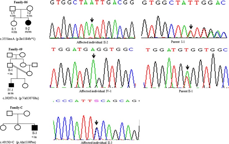
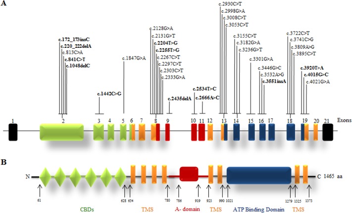
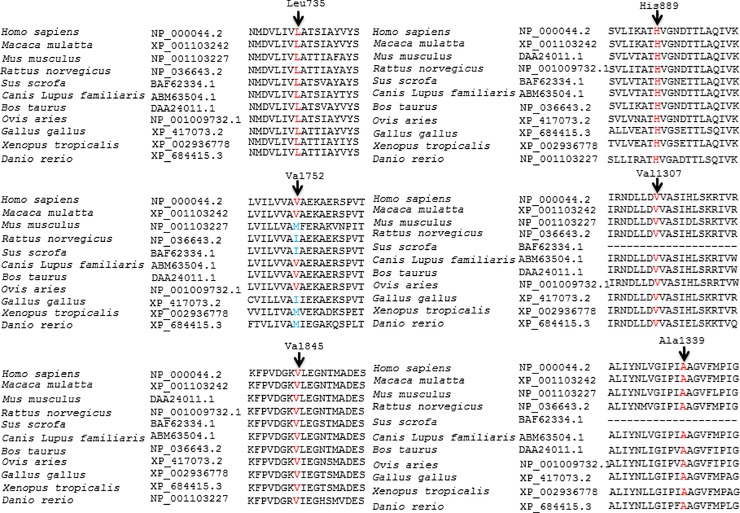
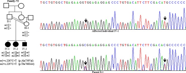
Wilson disease (WD) is an autosomal recessive disorder, characterized by excessive deposition of copper in various parts of the body, mainly in the liver and brain. It is caused by mutations in ATP7B. We report here the genetic analysis of 102 WD families from a south Indian population. Thirty-six different ATP7B mutations, including 13 novel ones [p.Ala58fs*19, p.Lys74fs*9, p.Gln281*, p.Pro350fs*12, p.Ser481*, p.Leu735Arg, p.Val752Gly, p.Asn812fs*2, p.Val845Ala, p.His889Pro, p.Ile1184fs*1, p.Val1307Glu and p.Ala1339Pro], were identified in 76/102 families. Interestingly, the mutation analysis of affected individuals in two families identified two different homozygous mutations in each family, and thus each affected individual from these families harbored two mutations in each ATP7B allele. Of 36 mutations, 28 were missense, thus making them the most prevalent mutations identified in the present study. Nonsense, insertion and deletion represented 3/36, 2/36 and 3/36 mutations, respectively. The haplotype analysis suggested founder effects for all the 14 recurrent mutations. Our study thus expands the mutational landscape of ATP7B with a total number of 758 mutations. The mutations identified during the present study will facilitate carrier and pre-symptomatic detection, and prenatal genetic diagnosis in affected families.


Conflict of interest statement

The authors have declared that no competing interests exist.

Figures

Fig 1
Fig 1. DNA sequence analysis of individuals from family-90.
(Upper panel) Sequencing chromatograms from the affected individual IV-1 and the parent III-1 showing c.172_173insC mutation. Arrows mark the insertion of C in a homozygous state in the affected individual IV-1 and in a heterozygous state in the parent III-1. (Lower panel) Sequencing chromatograms from the affected individual IV-1 and the parent III-1 showing c.3741C>G mutation. Arrows mark the C>G change in a homozygous state in the affected individual IV-1 and in a heterozygous state in the parent III-1. + denotes the wild-type allele. m1 and m2 denote different mutations.
Fig 2
Fig 2. DNA sequence analysis of individuals from family-128 and family-31.
(Upper panel) Sequencing chromatogram of the affected individual IV-5 from family-128. Arrow marks the deletion of A residue in a homozygous state. (Lower panel) Sequencing chromatograms of the affected individual II-3 and the parent I-1 from family-31. Arrows mark the C>T change in a homozygous state in the affected individual II-3 and in a heterozygous state in the parent I-1. + and m denote the wild-type and mutant alleles, respectively.
Fig 3
Fig 3. DNA sequence analysis of individuals from family-60 and family-506.
(Upper panel) Sequencing chromatograms from the affected individual V-1 and parent IV-1 from family-60. Arrows mark the deletion of the C residue in a homozygous state in the affected individual V-1 and in a heterozygous state in the parent IV-1. (Lower panel) Sequencing chromatograms from the affected individual II-1 and parent I-1 from family-506. Arrows mark the C>G change in a heterozygous state in the affected individual II-1 and parent I-1. + and m denote the wild type and mutant alleles, respectively.
Fig 4
Fig 4. DNA sequence analysis of individuals from family-M and family-41B.
(Upper panel) Sequencing chromatograms from the affected individual II-1 and parent I-1 from the family-M. Arrows mark the T>G change in a homozygous state in the affected individual II-1 and in a heterozygous state in the parent I-1. (Middle panel) Sequencing chromatogram from the affected individual II-1 from family-41B. Arrow marks the T>G change in a heterozygous state in the affected individual II-1. (Lower panel) Sequencing chromatogram from the affected individual II-1 from family-41B. Arrow marks the G>A change in a heterozygous state in the affected individual II-1. + denotes the wild type allele. m1 and m2 denote two different mutations.
Fig 5
Fig 5. DNA sequence analysis of individuals from family-77 and family-80.
(Upper panel) Sequencing chromatograms from the affected individual II-2 and parent I-1 from family-77. Arrows mark the deletion of the A residue in the affected individual II-2 and parent I-1 in a heterozygous state. (Lower panel) Sequencing chromatograms from the affected individual IV-1 and the parent II-2 from family-80. Arrows mark the T>C change in a homozygous state in the affected individual IV-1 and in a heterozygous state in the parent II-2. + and m denote the wild type and mutant alleles, respectively.
Fig 6
Fig 6. DNA sequence analysis of individuals from family-123.
(Upper panel) Sequencing chromatogram from the affected individual II-2 from family-123. Arrow marks the A>C change in a heterozygous state in the affected individual II-2. (Lower panel) Sequencing chromatogram from the affected individual II-2 and the parent I-2 from family-123. Arrows mark the C>T change in a heterozygous state in the affected individual II-2 and parent I-2. + denotes the wild type allele. m1 and m2 denote different mutations.
Fig 7
Fig 7. DNA sequence analysis of individuals from family-86, family-49 and family-C.
(Upper panel) Sequencing chromatograms from the affected individual II-2 and the parent I-1 from family-86. Arrows mark the insertion of the A residue in a homozygous state in the affected individual II-2 and in a heterozygous state in the parent I-1. (Middle panel) Sequencing chromatograms of the affected individual IV-1 and the parent II-1 from family-49. Arrows mark the T>A change in a homozygous state in the affected individual IV-1 and in a heterozygous state in the parent II-1. (Lower panel) Sequencing chromatogram from the affected individual II-3 from family-C. Arrow marks the G>C change in a heterozygous state in the affected individual II-3. + and m denote the wild type and mutant alleles, respectively.
Fig 8
Fig 8. Mutation landscape of the ATP7B gene and protein.
(A) The intron-exon structure of the gene. The novel mutations are shown in bold. (B) Different domains of the protein. Abbreviations: aa; amino acid; CBDs, copper binding domains; TMS, transmembrane segment and A-domain (actuator domain). The numbers refer to amino acid positions.
Fig 9
Fig 9. Conservation of the amino acid residues across different species in ATP7B.
Arrows mark the conservation of amino acid residues Leu735, Val752, Val845, His889, Val1307, and Ala1339 across different species. GenBank accession numbers of ATP7B are also given.
Fig 10
Fig 10. DNA sequence analysis of individuals from family-72.
Sequencing chromatograms from the affected individual IV-1 and parent II-1. Arrows mark two different C>T changes in a homozygous state in the affected individual IV-1 and in a heterozygous state in the parent II-1. + denotes the wild-type allele. m1 and m2 denote two different mutant alleles.

References

    1. Ala A, Borjigin J, Rochwarger A, Schilsky M. Wilson disease in septuagenarian siblings: raising the bar for diagnosis. Hepatology. 2005;4: 668–70. - PubMed
    1. Perri RE, Hahn SH, Ferber MJ, Kamath PS. Wilson disease-keeping the bar for diagnosis raised. Hepatology. 2005;42: 974. - PubMed
    1. Wiernicka A, Dądalski M, Jańczyk W, Kamińska D, Naorniakowska M, Hüsing-Kabar A, et al. Early onset of Wilson disease: diagnostic challenges. J. Pediatr. Gastroenterol. Nutr. 2017; 65: 555–560. 10.1097/MPG.0000000000001700 - DOI - PubMed
    1. Bull PC, Thomas GR, Rommens JM, Forbes JR, Cox DW. The Wilson disease gene is a putative copper transporting P-type ATPase similar to the Menkes gene. Nat. Genet. 1993;5: 327–337. 10.1038/ng1293-327 - DOI - PubMed
    1. Tanzi RE, Petrukhin K, Chernov I, Pellequer JL, Wasco W, Ross B et al. The Wilson disease gene is a copper transporting ATPase with homology to the Menkes disease gene. Nat. Genet. 1993;5: 344–350. 10.1038/ng1293-344 - DOI - PubMed

Publication types

Substances