. 2019 May 4;8(5):414.
doi: 10.3390/cells8050414.

Onset of Telomere Dysfunction and Fusions in Human Ovarian Carcinoma

Affiliations

Onset of Telomere Dysfunction and Fusions in Human Ovarian Carcinoma

Nazmul Huda et al. Cells. .

Abstract

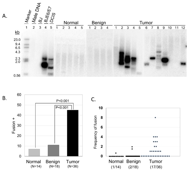
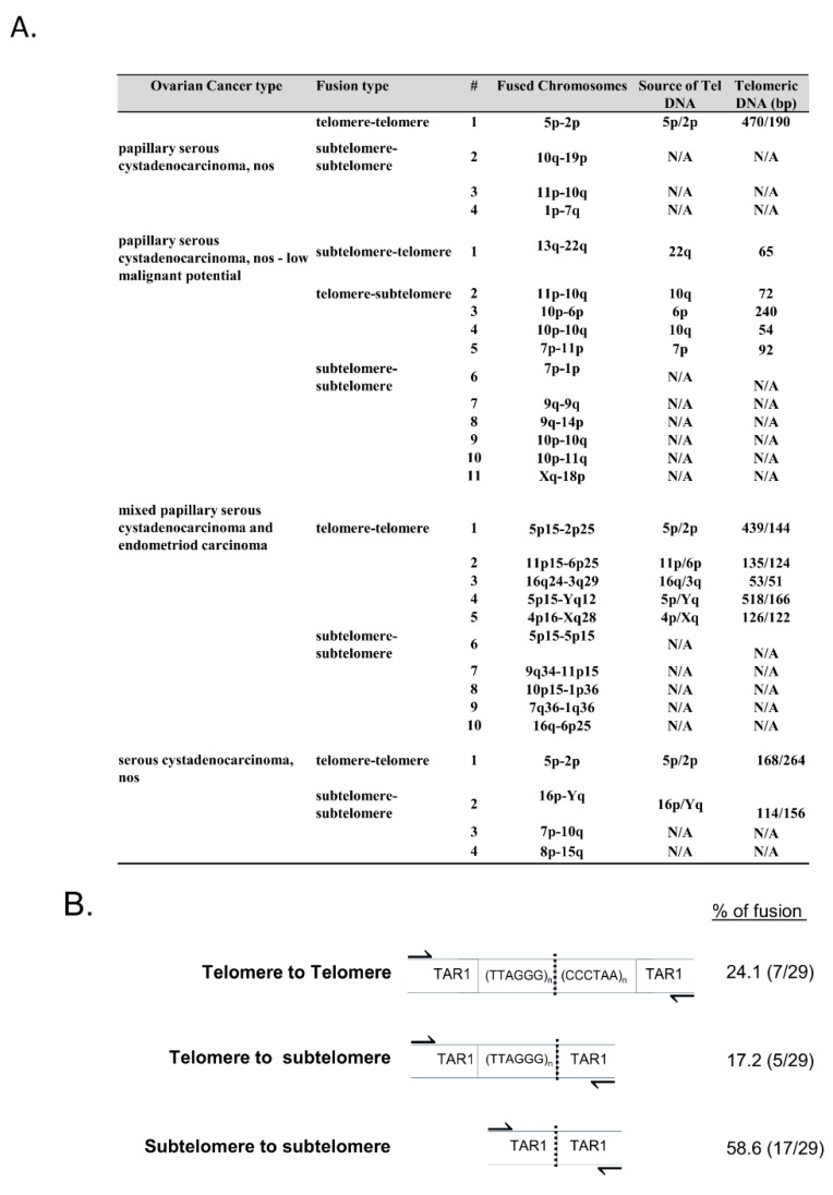
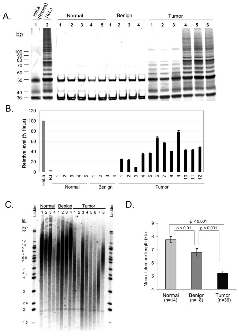
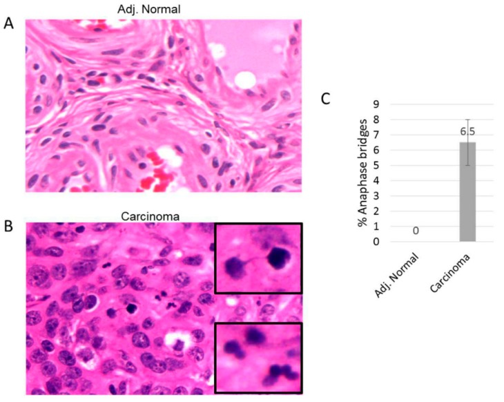
Telomere dysfunction has been strongly implicated in the initiation of genomic instability and is suspected to be an early event in the carcinogenesis of human solid tumors. Recent findings have established the presence of telomere fusions in human breast and prostate malignancies; however, the onset of this genomic instability mechanism during progression of other solid cancers is not well understood. Herein, we explored telomere dynamics in patient-derived epithelial ovarian cancers (OC), a malignancy characterized by multiple distinct subtypes, extensive molecular heterogeneity, and widespread genomic instability. We discovered a high frequency of telomere fusions in ovarian tumor tissues; however, limited telomere fusions were detected in normal adjacent tissues or benign ovarian samples. In addition, we found relatively high levels of both telomerase activity and hTERT expression, along with anaphase bridges in tumor tissues, which were notably absent in adjacent normal ovarian tissues and benign lesions. These results suggest that telomere dysfunction may occur early in ovarian carcinogenesis and, importantly, that it may play a critical role in the initiation and progression of the disease. Recognizing telomere dysfunction as a pervasive feature of this heterogeneous malignancy may facilitate the future development of novel diagnostic tools and improved methods of disease monitoring and treatment.

Keywords: genomic instability; ovarian carcinoma; telomerase; telomere; telomere dysfunction.


Conflict of interest statement

The authors declare no conflict of interest.

Figures

Figure 1
Figure 1
Telomere fusions in the ovarian tissue samples. (A) Southern blot analysis using 32P-labled [TTAGGG]4 probe. Five representative samples are shown for normal and benign, and 12 for tumor tissue samples. Normal male DNA and BJ foreskin fibroblast cells are used as negative controls, while BJ cells expressing the human papillomavirus type 16 E6/E7 oncoproteins (BJ E6/E7) DNA and ductal carcinoma in situ (DCIS) DNA are used as positive controls. (B) The percentage of fusion-positive samples. Standard t-test analysis is shown. (C) Frequency of telomere fusions where each dot represented one individual with the number of telomere fusion Southern bands per 500ng genomic DNA from five TAR-Fusion PCRs.
Figure 2
Figure 2
Telomere length and fusion analysis. (A) Each tumor tissue sample was evaluated for telomere length using qPCR. Each sample’s fusion type, categorization of fused chromosomes, and telomeric DNA (bp) length is shown. (B) Breakdown of the types of telomere fusion found in the above samples.
Figure 3
Figure 3
hTERT, telomerase, and telomere length profiles of ovarian carcinoma tissue samples. (A) Representative samples with telomerase activity in ovarian carcinoma. Fresh lysates of HeLa cells were used as positive control for telomerase activity while heat treated HeLa cells with inactivated telomerase serve as negative control. (B) Representative samples of hTERT expression levels in ovarian carcinoma quantified. HeLa and BJ cells were used as positive and negative controls respectively. (C) Representative Southern Blot of telomere length comparing normal, benign and tumor ovarian tissue. (D) Quantified mean telomere length (kb) of normal, benign and tumor tissue samples (mean ± SE). Standard t-test analysis is shown.
Figure 4
Figure 4
Anaphase bridge analysis in ovarian carcinoma. Tissue sections are stained with Hematoxylin and Eosin (H&E). (A) and (B) are representative adjacent tissue and ovarian carcinoma tissue sections, respectively. (C) The percent of anaphase bridges was calculated from the total anapases within each ovarian carcinoma.

References

    1. Palm W., Lange T.d. How Shelterin Protects Mammalian Telomeres. Annu. Rev. Genet. 2008;42:301–334. doi: 10.1146/annurev.genet.41.110306.130350. - DOI - PubMed
    1. Blackburn E.H., Epel E.S., Lin J. Human telomere biology: A contributory and interactive factor in aging, disease risks, and protection. Science. 2015;350:1193–1198. doi: 10.1126/science.aab3389. - DOI - PubMed
    1. Watson J.D. Origin of Cancatemeric T7 DNA. Nat. New Biol. 1972;239 doi: 10.1038/newbio239197a0. - DOI - PubMed
    1. Harley C.B., Futcher A.B., Greider C.W. Telomeres Shorten during Ageing of Human Fibroblasts. Nature. 1990;345:12. doi: 10.1038/345458a0. - DOI - PubMed
    1. O’Sullivan R.J., Karlseder J. Telomeres: Protecting chromosomes against genome instability. Nat. Rev. Mol. Cell Biol. 2010;11:171–181. doi: 10.1038/nrm2848. - DOI - PMC - PubMed