. 2019 Aug 1;25(15):4644-4655.
doi: 10.1158/1078-0432.CCR-19-0173. Epub 2019 May 6.

The Immune Microenvironment in Hormone Receptor-Positive Breast Cancer Before and After Preoperative Chemotherapy

Affiliations

The Immune Microenvironment in Hormone Receptor-Positive Breast Cancer Before and After Preoperative Chemotherapy

Adrienne G Waks et al. Clin Cancer Res. .

Abstract

Purpose: Hormone receptor-positive/HER2-negative (HR+/HER2-) breast cancer is associated with low levels of stromal tumor-infiltrating lymphocytes (sTIL) and PD-L1, and demonstrates poor responses to checkpoint inhibitor therapy. Evaluating the effect of standard chemotherapy on the immune microenvironment may suggest new opportunities for immunotherapy-based approaches to treating HR+/HER2- breast tumors.

Experimental design: HR+/HER2- breast tumors were analyzed before and after neoadjuvant chemotherapy. sTIL were assessed histologically; CD8+ cells, CD68+ cells, and PD-L1 staining were assessed immunohistochemically; whole transcriptome sequencing and panel RNA expression analysis (NanoString) were performed.

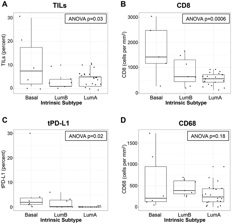
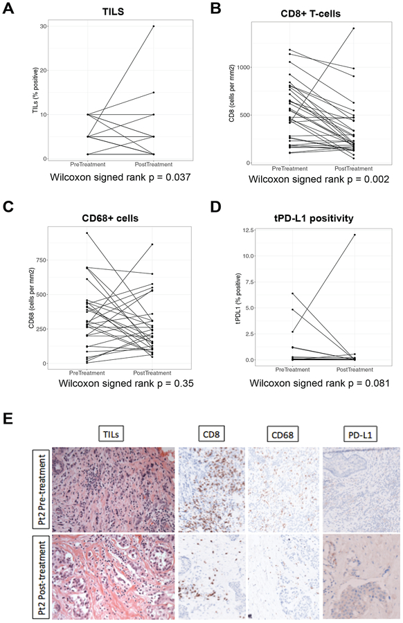
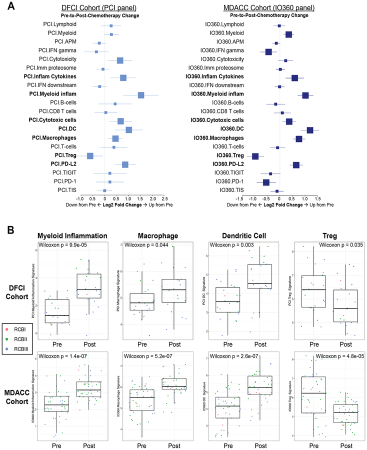
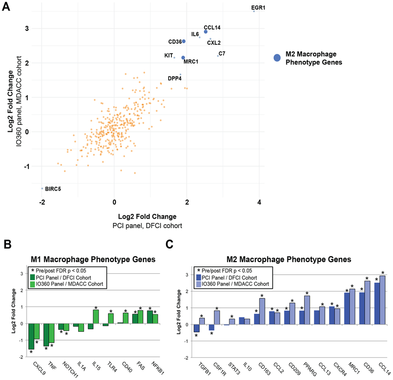
Results: Ninety-six patients were analyzed from two cohorts (n = 55, Dana-Farber cohort; n = 41, MD Anderson cohort). sTIL, CD8, and PD-L1 on tumor cells were higher in tumors with basal PAM50 intrinsic subtype. Higher levels of tissue-based lymphocyte (sTIL, CD8, PD-L1) and macrophage (CD68) markers, as well as gene expression markers of lymphocyte or macrophage phenotypes (NanoString or CIBERSORT), correlated with favorable response to neoadjuvant chemotherapy, but not with improved distant metastasis-free survival in these cohorts or a large gene expression dataset (N = 302). In paired pre-/postchemotherapy samples, sTIL and CD8+ cells were significantly decreased after treatment, whereas expression analyses (NanoString) demonstrated significant increase of multiple myeloid signatures. Single gene expression implicated increased expression of immunosuppressive (M2-like) macrophage-specific genes after chemotherapy.

Conclusions: The immune microenvironment of HR+/HER2- tumors differs according to tumor biology. This cohort of paired pre-/postchemotherapy samples suggests a critical role for immunosuppressive macrophage expansion in residual disease. The role of macrophages in chemoresistance should be explored, and further evaluation of macrophage-targeting therapy is warranted.


Figures

Figure 1.
Figure 1.. Tissue-based immune biomarkers and tumor intrinsic subtype.
In the DFCI cohort, stromal tumor-infiltrating lymphocytes (TILs; A) were evaluated histologically, and CD8+ cells (B), tumor cell PD-L1 expression (tPD-L1; C), and CD68+ cells (D) were stained immunohistochemically on full slide sections from treatment-naïve breast tumors. The distribution of each biomarker was assessed according to tumor intrinsic subtype (Luminal A, Luminal B, or Basal-like) based on the PAM50 gene signature. Each boxplot represents the 25th to 75th percentile with the median indicated as the central line and whiskers indicating 1.5 × interquartile range.
Figure 2.
Figure 2.. Gene-expression based biomarker correlation with chemotherapy response at surgery and long-term breast cancer outcomes.
Macrophage signature in the DFCI cohort was calculated using NanoString PanCancer Immune (PCI) Panel, and correlation with RCB status was assessed (A). M1-like macrophage inferred proportion by CIBERSORT and MDSC signature score(43) was calculated from microarray data from our prior meta-analysis of HR+/HER2− tumors treated with neoadjuvant chemotherapy(17), and correlation with RCB status was assessed (B-C). For A-C, due to a paucity of complete responses, pCR (RCB 0) and RCB-I were grouped together. Each boxplot represents the 25th to 75th percentile with the median indicated as the central line and whiskers indicating 1.5 × interquartile range. Association of M1-like macrophage or CD8 cell proportion by CIBERSORT with distant metastasis-free survival was assessed (D-E).
Figure 3.
Figure 3.. The tissue-based immune microenvironment before and after chemotherapy (DFCI cohort).
Tissue-based immune biomarkers were scored on paired slides from pre- versus post-preoperative therapy with dose-dense adriamycin/cyclophosphamide plus bevacizumab (A-D). Histology images (400×) from a patient with pre-to-post-treatment decrease in stromal tumor-infiltrating lymphocytes (sTIL), CD8, and CD68 (10% to 5%, 842 cells/mm2 to 206 cells/mm2, and 414 cells/mm2 to 145 cells/mm2 pre- to post-treatment, respectively) are shown. PD-L1 expression on tumor (tPD-L1) and stromal cells was 0% both pre- and post-treatment for this patient (E).
Figure 4.
Figure 4.. NanoString gene expression signatures before and after chemotherapy.
Immune gene expression signatures by PanCancer Immune Profiling Panel (PCI; DFCI cohort) or the PanCancer IO 360 Profiling Panel (IO360; MD Anderson cohort) were assessed pre- and post-neoadjuvant chemotherapy. Signatures that changed significantly in both cohorts are indicated in bold (A). Individual box-and-whisker plots highlight signatures of particular interest in both cohorts, with associated statistical test shown for significant change and points colored according to RCB status of each individual patient (B). Each boxplot represents the 25th to 75th percentile with the median indicated as the central line and whiskers indicating 1.5 × interquartile range. Abbreviations: MDACC, MD Anderson Cancer Center.
Figure 5.
Figure 5.. NanoString individual gene expression before and after chemotherapy.
Individual gene expression by PanCancer Immune Profiling Panel (PCI; DFCI cohort) or the PanCancer IO 360 Profiling Panel (IO360; MD Anderson cohort) were assessed pre- and post-neoadjuvant chemotherapy. Genes were ranked by fold-change (averaged across both cohorts) and the top 10 genes by absolute value of average fold-change, with false discovery rate p-value <0.05 in both cohorts, are labeled. Blue dots indicate genes that relate to M2-like macrophage phenotype; there were no genes in the top 10 that related to M1-like macrophage phenotype (A). Individual genes associated with M1-like versus M2-like phenotype were hand curated after comprehensive literature search (Table S3) and plotted according to log2 fold change (B). Abbreviations: MDACC, MD Anderson Cancer Center.

References

    1. Howlader N, Altekruse SF, Li CI, Chen VW, Clarke CA, Ries LA, et al. US incidence of breast cancer subtypes defined by joint hormone receptor and HER2 status. Journal of the National Cancer Institute 2014;106(5) doi 10.1093/jnci/dju055. - DOI - PMC - PubMed
    1. Issa-Nummer Y, Darb-Esfahani S, Loibl S, Kunz G, Nekljudova V, Schrader I, et al. Prospective validation of immunological infiltrate for prediction of response to neoadjuvant chemotherapy in HER2−negative breast cancer--a substudy of the neoadjuvant GeparQuinto trial. PloS one 2013;8(12):e79775 doi 10.1371/journal.pone.0079775. - DOI - PMC - PubMed
    1. Muenst S, Schaerli AR, Gao F, Daster S, Trella E, Droeser RA, et al. Expression of programmed death ligand 1 (PD-L1) is associated with poor prognosis in human breast cancer. Breast cancer research and treatment 2014;146(1):15–24 doi 10.1007/s10549-014-2988-5. - DOI - PMC - PubMed
    1. Vidula N, Yau C, Goga A, Rugo HS. Programmed cell death 1 (PD-1) receptor and prograamed death ligand 1 (PD-L1) expression in primary breast cancer (BC); correlations with clinical characteristics and patient outcomes. Journal of clinical oncology : official journal of the American Society of Clinical Oncology 2015;33:suppl; abstr 1090.
    1. Ghebeh H, Mohammed S, Al-Omair A, Qattan A, Lehe C, Al-Qudaihi G, et al. The B7-H1 (PD-L1) T lymphocyte-inhibitory molecule is expressed in breast cancer patients with infiltrating ductal carcinoma: correlation with important high-risk prognostic factors. Neoplasia 2006;8(3):190–8 doi 10.1593/neo.05733. - DOI - PMC - PubMed

Publication types

MeSH terms