

Brain ventricular volume changes induced by long-duration spaceflight
- PMID: 31061119
- PMCID: PMC6535034
- DOI: 10.1073/pnas.1820354116
Brain ventricular volume changes induced by long-duration spaceflight
Abstract
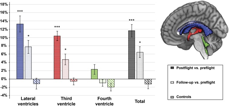
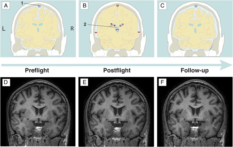
Long-duration spaceflight induces detrimental changes in human physiology. Its residual effects and mechanisms remain unclear. We prospectively investigated the changes in cerebrospinal fluid (CSF) volume of the brain ventricular regions in space crew by means of a region of interest analysis on structural brain scans. Cosmonaut MRI data were investigated preflight (n = 11), postflight (n = 11), and at long-term follow-up 7 mo after landing (n = 7). Post hoc analyses revealed a significant difference between preflight and postflight values for all supratentorial ventricular structures, i.e., lateral ventricle (mean % change ± SE = 13.3 ± 1.9), third ventricle (mean % change ± SE = 10.4 ± 1.1), and the total ventricular volume (mean % change ± SE = 11.6 ± 1.5) (all P < 0.0001), with higher volumes at postflight. At follow-up, these structures did not quite reach baseline levels, with still residual increases in volume for the lateral ventricle (mean % change ± SE = 7.7 ± 1.6; P = 0.0009), the third ventricle (mean % change ± SE = 4.7 ± 1.3; P = 0.0063), and the total ventricular volume (mean % change ± SE = 6.4 ± 1.3; P = 0.0008). This spatiotemporal pattern of CSF compartment enlargement and recovery points to a reduced CSF resorption in microgravity as the underlying cause. Our results warrant more detailed and longer longitudinal follow-up. The clinical impact of our findings on the long-term cosmonauts' health and their relation to ocular changes reported in space travelers requires further prospective studies.
Keywords: CSF; brain; microgravity; spaceflight; ventricles.
Copyright © 2019 the Author(s). Published by PNAS.
Conflict of interest statement
The authors declare no conflict of interest.
Figures
Comment in
-
The buffering capacity of the brain and optic nerve against spaceflight-associated neuro-ocular syndrome.Proc Natl Acad Sci U S A. 2019 Aug 6;116(32):15770-15771. doi: 10.1073/pnas.1908865116. Epub 2019 Jul 30. Proc Natl Acad Sci U S A. 2019. PMID: 31363044 Free PMC article. No abstract available.
-
Reply to Wostyn et al.: Investigating the spaceflight-associated neuro-ocular syndrome and the human brain in lockstep.Proc Natl Acad Sci U S A. 2019 Aug 6;116(32):15772-15773. doi: 10.1073/pnas.1909828116. Epub 2019 Jul 30. Proc Natl Acad Sci U S A. 2019. PMID: 31363045 Free PMC article. No abstract available.
-
Reply to Ludwig et al.: A potential mechanism for intracranial cerebrospinal fluid accumulation during long-duration spaceflight.Proc Natl Acad Sci U S A. 2019 Oct 8;116(41):20265-20266. doi: 10.1073/pnas.1913041116. Epub 2019 Sep 17. Proc Natl Acad Sci U S A. 2019. PMID: 31530727 Free PMC article. No abstract available.
-
Breathing drives CSF: Impact on spaceflight disease and hydrocephalus.Proc Natl Acad Sci U S A. 2019 Oct 8;116(41):20263-20264. doi: 10.1073/pnas.1910305116. Epub 2019 Sep 17. Proc Natl Acad Sci U S A. 2019. PMID: 31530729 Free PMC article. No abstract available.
References
-
- Clément G. (2011) Fundamentals of Space Medicine (Springer, Dordrecht, The Netherlands: ), 2nd Ed.
-
- Tarver W, Otto C (2012) NASA’s spaceflight visual impairment intracranial pressure (VIIP) risk: Clinical correlations and pathophysiology. Aerospace Medicine Grand Rounds (NASA, Washington, DC: ).
-
- Mader TH, et al. (2011) Optic disc edema, globe flattening, choroidal folds, and hyperopic shifts observed in astronauts after long-duration space flight. Ophthalmology 118:2058–2069. - PubMed
Publication types
MeSH terms
LinkOut - more resources
Full Text Sources
