. 2019 Jun 20;87(7):e00295-19.
doi: 10.1128/IAI.00295-19. Print 2019 Jul.

Citrobacter rodentium Induces Tissue-Resident Memory CD4+ T Cells

Affiliations

Citrobacter rodentium Induces Tissue-Resident Memory CD4+ T Cells

S Bishu et al. Infect Immun. .

Abstract

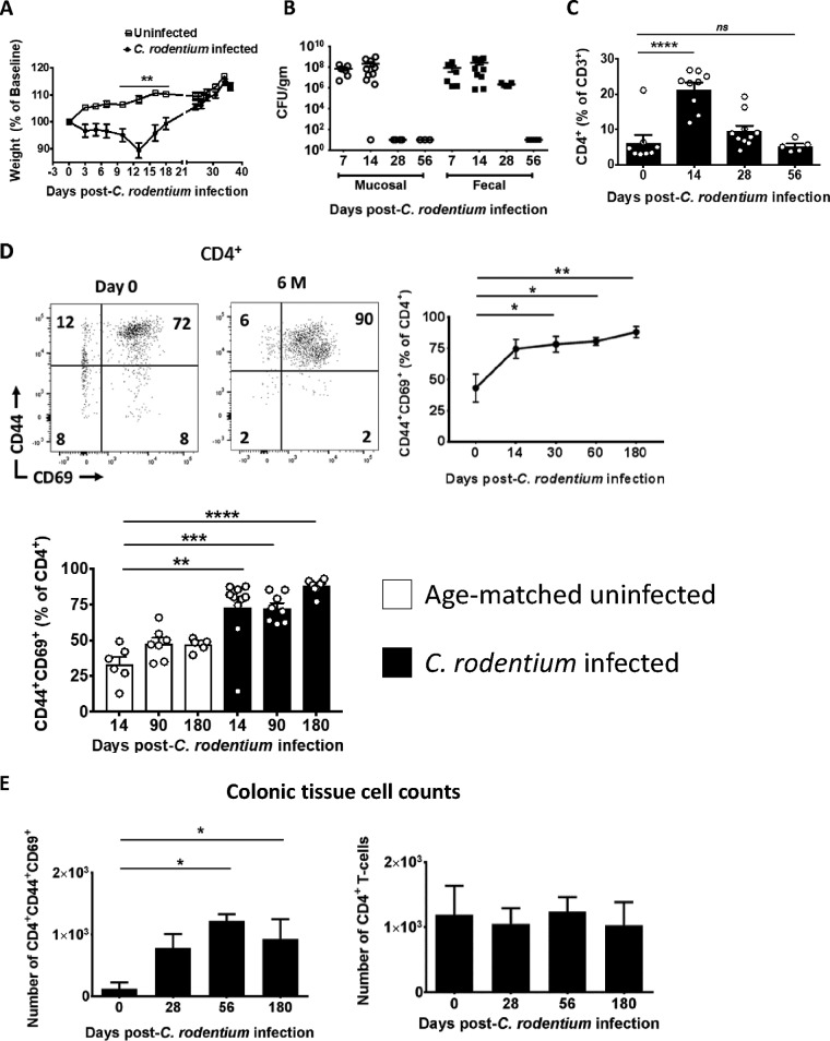
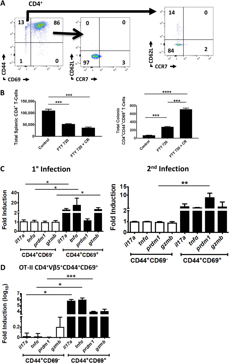
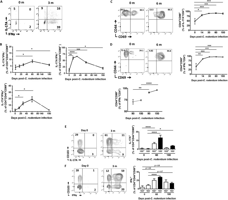
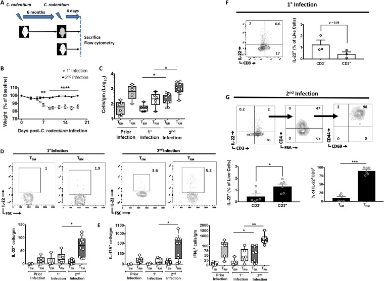
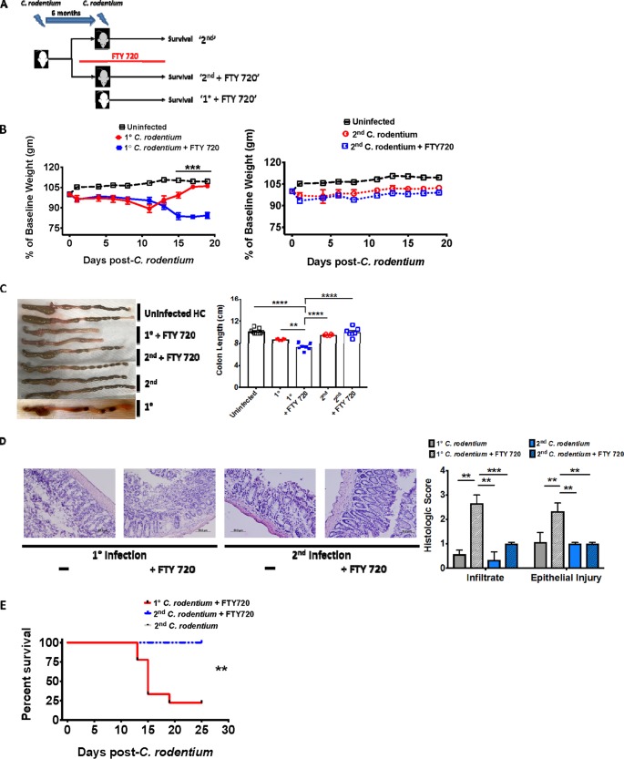
Tissue-resident memory T cells (TRM cells) are a novel population of tissue-restricted antigen-specific T cells. TRM cells are induced by pathogens and promote host defense against secondary infections. Although TRM cells cannot be detected in circulation, they are the major memory CD4+ and CD8+ T-cell population in tissues in mice and humans. Murine models of CD8+ TRM cells have shown that CD8+ TRM cells maintain tissue residency via CD69 and though tumor growth factor β-dependent induction of CD103. In contrast to CD8+ TRM cells, there are few models of CD4+ TRM cells. Thus, much less is known about the factors regulating the induction, maintenance, and host defense functions of CD4+ TRM cells. Citrobacter rodentium is known to induce IL-17+ and IL-22+ CD4+ T cells (Th17 and Th22 cells, respectively). Moreover, data from IL-22 reporter mice show that most IL-22+ cells in the colon 3 months after C. rodentium infection are CD4+ T cells. This collectively suggests that C. rodentium may induce CD4+ TRM cells. Here, we demonstrate that C. rodentium induces a population of IL-17A+ CD4+ T cells that are tissue restricted and antigen specific, thus meeting the criteria of CD4+ TRM cells. These cells expand and are a major source of IL-22 during secondary C. rodentium infection, even before the T-cell phase of the host response in primary infection. Finally, using FTY 720, which depletes circulating naive and effector T cells but not tissue-restricted T cells, we show that these CD4+ TRM cells can promote host defense.

Keywords: CD4+; Citrobacter; T cells; colitis; tissue-resident memory.


Figures

FIG 1
FIG 1
C. rodentium-induced alterations in the colonic CD4+ T-cell distribution are maintained over the long term. (A) Weight change of C. rodentium-infected and uninfected control mice. WT mice were orally gavaged with C. rodentium, and weight as a percentage of baseline was determined. (B) WT mice eradicate mucosal and luminal C. rodentium within 5 weeks. Bacterial load in colonic mucosa and colonic fecal pellets, expressed as CFU/g of intestine or stool, was determined at the indicated times. (C) The fraction of CD4+ T cells as a percentage of CD3+ T cells peaks by day 14. The fraction of CD4+ T cells as a percentage of CD3+ cells was determined by flow cytometry in the colon tissue of C. rodentium-infected mice at the indicated times. (D) Most mucosal CD4+ T cells exhibit a surface phenotype consistent with TRM cells after C. rodentium infection. Mononuclear cells were isolated from the colon of C. rodentium-infected mice at the indicated time points, and expression of CD44 and CD69 was determined on live CD3+ CD4+ T cells (top left panel; representative plot) and is quantitated relative to uninfected 6-week-old mice at day 0 (top right panel) and relative to age- and gender-matched uninfected mice (bottom left panel). (E) The total number of CD4+ CD44+ CD69+ T cells in the colon increases after C. rodentium infection. The total number of CD4+ CD44+ CD69+ T cells (left panel) and CD4+ T cells (right panel) in the colon was determined at the indicated times using flow cytometry normalized to colon weight (cells/g). Each time point consists of 5 to 10 mice (A to C) or 6 to 10 mice (D and E). *, P < 0.05; **, P < 0.01; ***, P < 0.001; ****, P < 0.0001, by parametric Student’s t test (A) or ANOVA (B to E).
FIG 2
FIG 2
C. rodentium-induced CD4+ CD44+ CD69+ cells are nonrecirculating and contain an antigen-specific subset. (A) C. rodentium-induced CD4+ CD44+ CD69+ T cells do not express lymph node homing markers. Expression of the lymph node homing markers CD62L (L-selectin) and CCR7 was determined on CD4+ CD44+ CD69+ and CD4+ CD44+ CD69 mucosal T cells 8 weeks after C. rodentium infection. (B) C. rodentium-induced CD4+ CD44+ CD69+ T cells do not recirculate. The total number of CD4+ T cells in the spleen (left panel) and CD4+ CD44+ CD69+ T cells in the colon (right panel) was enumerated with flow cytometry in uninfected WT mice (control) and mice administered FTY 720 with or without prior C. rodentium (CR) infection. (C) Mucosal CD4+ CD44+ CD69+ T cells contain a C. rodentium antigen-specific population over the long term after C. rodentium infection. Expression of genes associated with C. rodentium-induced Th17 cells was determined in the CD69+ and CD69 fractions of FACS-purified CD4+ CD44+ mucosal T cells in WT mice 8 weeks after primary infection (left panel) or 10 days after secondary infection (right panel). (D) Antigen-specific Th17 cells are contained in the CD4+ CD44+ CD69+ fraction in OT-II mice. CD4+ Vβ5+ CD44+ CD69+ colonic T cells were FACS purified from OT-II mice 10 days after inoculation with OVA-expressing C. rodentium, and expression of the selected genes was determined. Data are normalized to GAPDH and presented as fold induction over CD4+ CD44+ CD69 fractions for panels C and D. The experiment in panel A is representative of 6 mice. Each time point in panel B consists of 9 mice. Experiments in panels C and D were performed twice with 3 mice per trial (6 mice total). *, P < 0.5; **, P ≤ 0.01; ***, P ≤ 0.001; ****, P < 0.0001, by ANOVA.
FIG 3
FIG 3
C. rodentium-induced CD4+ TRM cells exhibit a Th1 and Th17 profile. (A) Mucosal CD4+ CD44+ CD69+ T cells exhibit a Th17 and Th1/17 signature over the long term after C. rodentium infection. Mucosal CD4+ CD44+ CD69+ T cells isolated from the colon of C. rodentium-infected mice at the indicated time points were stimulated with phorbol myristate acetate and ionomycin, and expression of IL-17A and of IFN-γ was determined. m, months. (B) Data from panel A are quantified and presented as IL-17A and IFN-γ single-positive (top left and right panels, respectively) and double-positive (bottom panel) CD4+ TRM cells. (C and D) Increasing fractions of IL-17A- (C) and IFN-γ-producing (D) CD4+ T cells express TRM markers after C. rodentium infection. Expression of CD44 and CD69 on IL-17A+ (C) and IFN-γ+ (D) mucosal CD4+ T cells from C. rodentium-infected mice was determined at baseline (0) and 6 months (6 m) relative to uninfected 6-week-old mice or relative to uninfected age-matched controls (D, bottom panel). The following gating schema was used: live lymphocytes→IL-17A+ (or IFN-γ+)→CD3+→CD4+→CD44 versus CD69. FACS plots (left panels) and quantitated data (right panels and bottom panel of panel D) are presented. (E and F) C. rodentium-induced CD4+ TRM cells produce IL-17 and IFN-γ independently of CD103 expression. Coordinated expression of CD103 and IL-17A (E) or CD103 and IFN-γ (F) was determined in CD4+ CD44+ CD69+ mucosal T cells at the indicated time points postinfection and quantified (right panels). Panel A is a representative plot, while 6 to 9 mice (B and C) or 6 to 11 mice (E and F) are presented for each time point. *, P < 0.5; **, P ≤ 0.01; ***, P ≤ 0.001; ****, P < 0.0001, by ANOVA.
FIG 4
FIG 4
C. rodentium-induced CD4+ TRM cells are reactivated and are a source of IL-22 during secondary infections. (A) The experimental scheme. WT mice that had recovered from C. rodentium infection 6 months before were reinfected and sacrificed 4 days postinfection (dark gray). Age- and gender-matched mice that had primary infection served as controls (white). (B) Weight loss for mice postinfection. (C) CD4+ TRM cell compartment expands with secondary infection. The total number of colonic CD4+ CD44+ CD69 (TEM) and CD4+ CD44+ CD69+ (TRM) T cells was enumerated with flow cytometry 4 days after primary or secondary infection of mice per the schema in panel A. The number of TEM and TRM cells was also quantified in age-matched mice that had cleared C. rodentium infection 6 months previously (Prior Infection) as an additional control. (D and E) IL-22+ (D) and IL-17+ (left panel) and IFN-γ+ (right panel) (E) CD4+ TRM cell compartments expand with secondary infection. (D) Representative FACS plots (top panels) and quantified total numbers of IL-22+ (D) and IL-17A+ (E, left panel) and IFN-γ+ (E, right panel) CD4+ CD44+ CD69 (TEM) and CD4+ CD44+ CD69+ (TRM) T cells were enumerated with flow cytometry 4 days after primary or secondary infection of mice per the schema in panel A. The number of IL-22+, IL-17A+, and IFN-γ+ TEM and TRM cells was also quantified in age-matched mice that had cleared C. rodentium infection 6 months before (Prior Infection). (F) Most IL-22+ cells early during primary infection are CD3. Coordinated expression of IL-22 and CD3 was determined in primary-infection mice day 4 postinfection. (G) T cells with a TRM phenotype are a source of IL-22 early during secondary infection. Sequential expression of CD4 (top row, middle panel) and CD44 and CD69 (top row, right panel) was determined on live IL-22+ CD3+ mucosal cells (top row, left panel). Cumulative data are quantified (bottom row). Data are expressed as cells per gram of colon (C to E). Experiments in panels B to E and G comprise 6 to 10 mice per group. The experiment in panel F is representative of an experiment with 3 mice (performed twice). *, P < 0.5; **, P ≤ 0.01; ***, P ≤ 0.001, by the parametric Student t test (B, F, and G) or ANOVA (C to E).
FIG 5
FIG 5
Circulating naive T cells are redundant for host defense during secondary C. rodentium infection. (A) The experimental scheme. WT mice that had recovered from primary C. rodentium infection 6 months previously were reinfected (gray) with or without FTY 720 and compared to primary-infection mice (white). FTY 720 was administered in primary-infection and a subset of secondary-infection mice 14 days prior to infection and continued for the duration of the experiment. (B, C, and D) Circulating naive T cells are redundant for host defense against secondary C. rodentium infection. (B) Weight loss in uninfected controls and primary-infection (left panel) and secondary-infection mice (right panel) with or without FTY 720. (C) Colon length in uninfected control and on day 15 after primary and secondary C. rodentium infection with or without FTY 720. (D) Hematoxylin-and-eosin-stained sections and quantified histologic score (bottom row) in primary-infection (left panel) and secondary-infection mice (right panel) with or without FTY 720 and primary-infection age- and gender-matched mice. (E) Noncirculating T cells promote host defense against secondary C. rodentium infections. Overall survival in primary-infection and secondary-infection mice with or without FTY 720. Experiments in panels B to E consist of 6 to 7 mice per group, repeated twice. Data from a representative experiment are shown in panels B to E. **, P < 0.01; ***, P ≤ 0.001; ****, P < 0.0001, by the parametric Student t test (B), ANOVA (C and D), or Kaplan-Meier method (E).

References

    1. Mueller SN, Mackay LK. 2016. Tissue-resident memory T cells: local specialists in immune defence. Nat Rev Immunol 16:79–89. doi: 10.1038/nri.2015.3. - DOI - PubMed
    1. Park CO, Kupper TS. 2015. The emerging role of resident memory T cells in protective immunity and inflammatory disease. Nat Med 21:688–697. doi: 10.1038/nm.3883. - DOI - PMC - PubMed
    1. Iijima N, Iwasaki A. 2014. T cell memory. A local macrophage chemokine network sustains protective tissue-resident memory CD4 T cells. Science 346:93–98. doi: 10.1126/science.1257530. - DOI - PMC - PubMed
    1. Zens KD, Chen JK, Farber DL. 2016. Vaccine-generated lung tissue-resident memory T cells provide heterosubtypic protection to influenza infection. JCI Insight 1:85832. doi: 10.1172/jci.insight.85832. - DOI - PMC - PubMed
    1. Shiow LR, Rosen DB, Brdickova N, Xu Y, An J, Lanier LL, Cyster JG, Matloubian M. 2006. CD69 acts downstream of interferon-alpha/beta to inhibit S1P1 and lymphocyte egress from lymphoid organs. Nature 440:540–544. doi: 10.1038/nature04606. - DOI - PubMed

Publication types