. 2019 May 6;9(1):6996.
doi: 10.1038/s41598-019-43407-z.

Comprehensive Analysis of Animal Models of Cardiovascular Disease using Multiscale X-Ray Phase Contrast Tomography

Affiliations

Comprehensive Analysis of Animal Models of Cardiovascular Disease using Multiscale X-Ray Phase Contrast Tomography

Hector Dejea et al. Sci Rep. .

Erratum in

Abstract

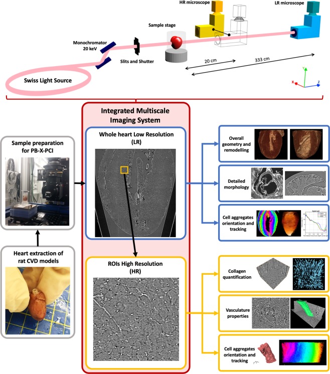
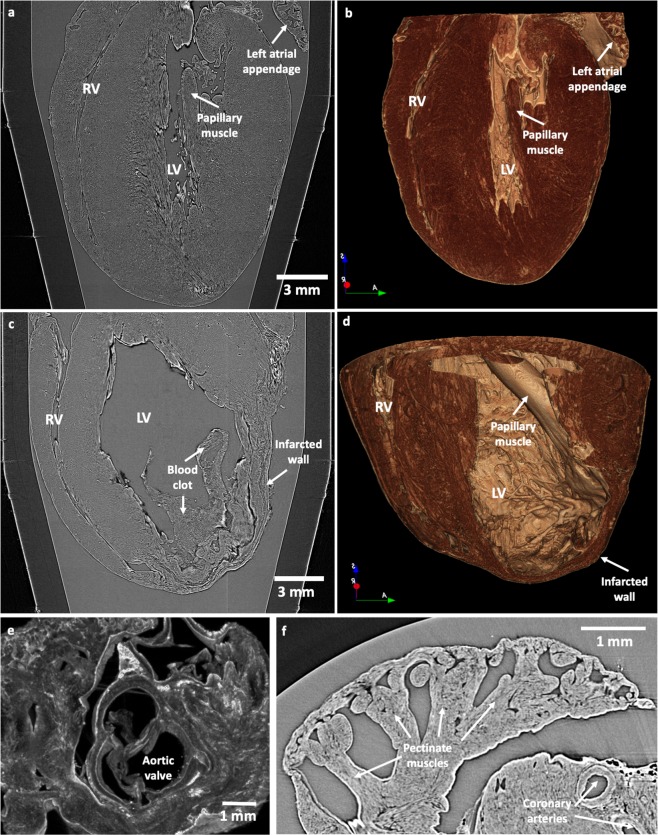
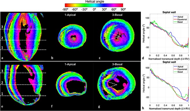
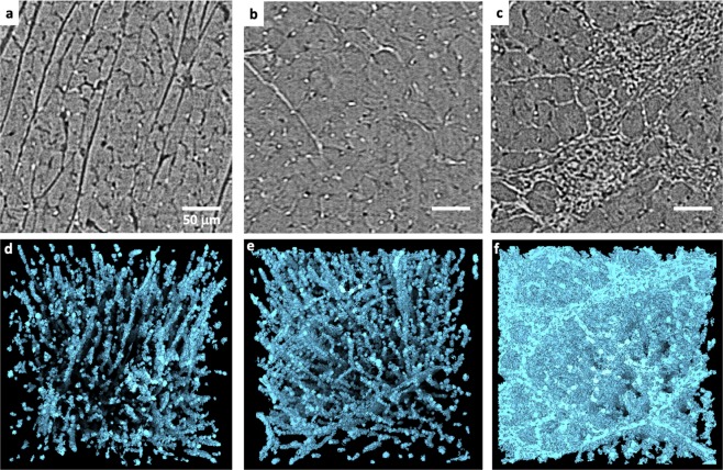
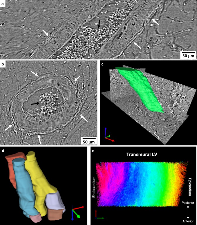
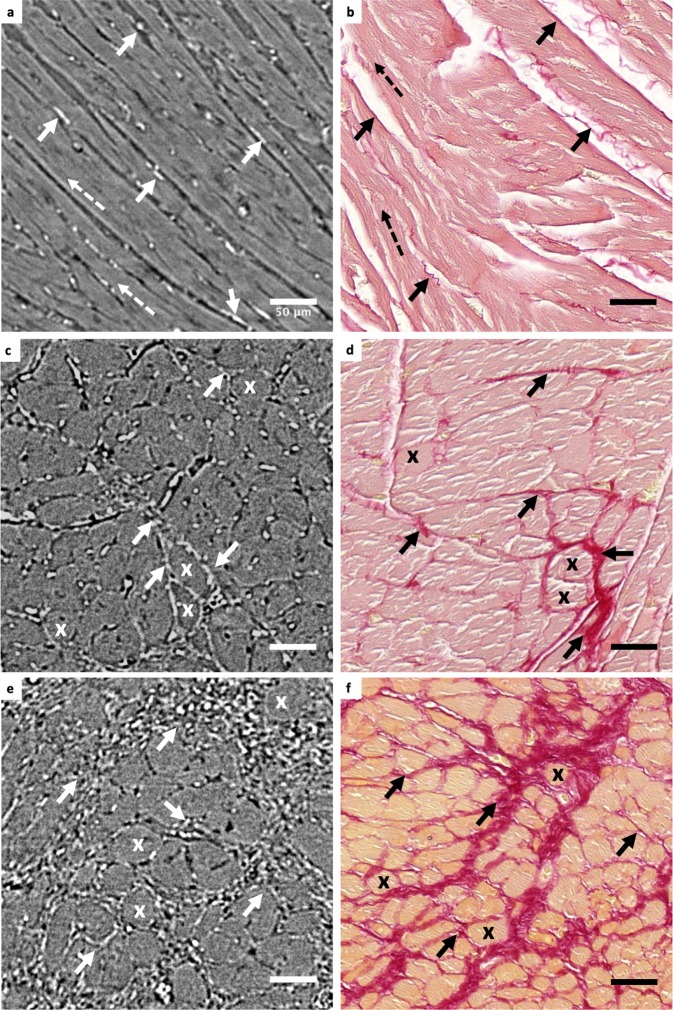
Cardiovascular diseases (CVDs) affect the myocardium and vasculature, inducing remodelling of the heart from cellular to whole organ level. To assess their impact at micro and macroscopic level, multi-resolution imaging techniques that provide high quality images without sample alteration and in 3D are necessary: requirements not fulfilled by most of current methods. In this paper, we take advantage of the non-destructive time-efficient 3D multiscale capabilities of synchrotron Propagation-based X-Ray Phase Contrast Imaging (PB-X-PCI) to study a wide range of cardiac tissue characteristics in one healthy and three different diseased rat models. With a dedicated image processing pipeline, PB-X-PCI images are analysed in order to show its capability to assess different cardiac tissue components at both macroscopic and microscopic levels. The presented technique evaluates in detail the overall cardiac morphology, myocyte aggregate orientation, vasculature changes, fibrosis formation and nearly single cell arrangement. Our results agree with conventional histology and literature. This study demonstrates that synchrotron PB-X-PCI, combined with image processing tools, is a powerful technique for multi-resolution structural investigation of the heart ex-vivo. Therefore, the proposed approach can improve the understanding of the multiscale remodelling processes occurring in CVDs, and the comprehensive and fast assessment of future interventional approaches.


Conflict of interest statement

The authors declare no competing interests.

Figures

Figure 1
Figure 1
Multiscale imaging and analysis of cardiac tissue. Diagram describing the processing pipeline and imaging setup of the methodology used for comprehensive cardiac tissue analysis.
Figure 2
Figure 2
Whole heart geometry and detailed morphology assessment by PB-X-PCI. (a) Illustrative longitudinal PB-X-PCI virtual cut of the WKY heart at 5.8 μm pixel size (LR). (b) 3D volume rendered image showing the detailed internal structure of both ventricles extracted from the PB-X-PCI dataset in (a). (c) Illustrative longitudinal PB-X-PCI virtual cut of the LAD heart at 5.8 μm pixel size (LR). (d) 3D volume rendered image of the detailed internal structure of both ventricles and the infarcted region, extracted from the PB-X-PCI dataset in (c). (e) 3D detailed visualization of the aortic valve and surrounding structures in the SHR heart. (f) PB-X-PCI image (reconstructed with Paganin’s method) of the pectinate muscles in the left atrial appendage, as well as large coronary arteries in the myocardial wall of the SHR heart.
Figure 3
Figure 3
Myocyte aggregates orientation quantification. For WKY (top) and LAD (bottom) hearts, respectively: Ventricular helical angle maps in (a,e) four chambers view, as well as in (b,f) apical and (c,g) basal cross sections slices (marked with dashed white lines in the four chambers views). Colour coded by helical angle values. (d,h) Transmural profile from left-side endocardium to right-side endocardium of helical angle in the septal wall (dashed triangular area highlighted in c and g) at three different apico-basal positions (marked with dashed white lines and numbers in the four chambers views: 1-apical, 2-equatorial, 3-basal).
Figure 4
Figure 4
Collagen segmentation in HR images. (ac) Representative PB-X-PCI image slices from 300 × 300 × 300 μm subvolumes in the left ventricular septum of the WKY, SHR and ISO hearts respectively. (df) 3D renderings of collagen segmentation in the same subvolumes, visually showing the increase in density and change in shape and distribution around the tissue.
Figure 5
Figure 5
Qualitative analysis of microstructural components of cardiac tissue. (a) Longitudinal and (b) cross section cuts of a representative coronary artery in the left ventricular (LV) wall of the SHR heart. The white arrows point at the collagen forming the adventitia layer around the vessel wall in the hypertensive rat. Black arrows indicate the presence of blood cells. (c) 3D rendering of a section of the visualised artery. (d) 3D rendering of a selected bundle of myocytes in the LV of the LAD heart. Colours were changed when branching was observed. (e) Fibre tracking in the HR LV transmural volume of the WKY heart. Image was colour coded by helical angle values as in Fig. 3.
Figure 6
Figure 6
Histological validation of PB-X-PCI. PB-X-PCI images from the apex of (a) WKY, (c) SHR and (e) ISO in comparison with (b,d,f) histological slides stained with Picrosirius red from the approximate same tissue region respectively, with collagen shown in red. Bold arrows indicate the presence of fibrosis, dashed arrows follow longitudinal myocyte direction and crosses mark cross sectional view of the myocytes in both image types.

References

    1. Burchfield Jana S., Xie Min, Hill Joseph A. Pathological Ventricular Remodeling. Circulation. 2013;128(4):388–400. doi: 10.1161/CIRCULATIONAHA.113.001878. - DOI - PMC - PubMed
    1. Cohn Jay N, Ferrari Roberto, Sharpe Norman. Cardiac remodeling—concepts and clinical implications: a consensus paper from an international forum on cardiac remodeling. Journal of the American College of Cardiology. 2000;35(3):569–582. doi: 10.1016/S0735-1097(99)00630-0. - DOI - PubMed
    1. Mozaffarian, D. et al. Heart disease and stroke statistics-2016 update a report from the American Heart Association. Circulation, 10.1161/CIR.0000000000000350 (2016). - PubMed
    1. Zaragoza Carlos, Gomez-Guerrero Carmen, Martin-Ventura Jose Luis, Blanco-Colio Luis, Lavin Begoña, Mallavia Beñat, Tarin Carlos, Mas Sebastian, Ortiz Alberto, Egido Jesus. Animal Models of Cardiovascular Diseases. Journal of Biomedicine and Biotechnology. 2011;2011:1–13. doi: 10.1155/2011/497841. - DOI - PMC - PubMed
    1. Engle Steven K., Jordan William H., Pritt Michael L., Chiang Alan Y., Davis Myrtle A., Zimmermann John L., Rudmann Daniel G., Heinz-Taheny Kathleen M., Irizarry Armando R., Yamamoto Yumi, Mendel David, Schultze A. Eric, Cornwell Paul D., Watson David E. Qualification of Cardiac Troponin I Concentration in Mouse Serum Using Isoproterenol and Implementation in Pharmacology Studies to Accelerate Drug Development. Toxicologic Pathology. 2009;37(5):617–628. doi: 10.1177/0192623309339502. - DOI - PubMed

Publication types

MeSH terms