. 2019 Aug;138(2):275-293.
doi: 10.1007/s00401-019-02021-z. Epub 2019 May 6.

RhoA regulates translation of the Nogo-A decoy SPARC in white matter-invading glioblastomas

Affiliations

RhoA regulates translation of the Nogo-A decoy SPARC in white matter-invading glioblastomas

Peter Wirthschaft et al. Acta Neuropathol. 2019 Aug.

Abstract

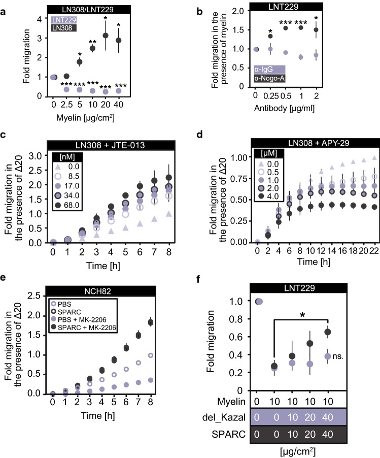
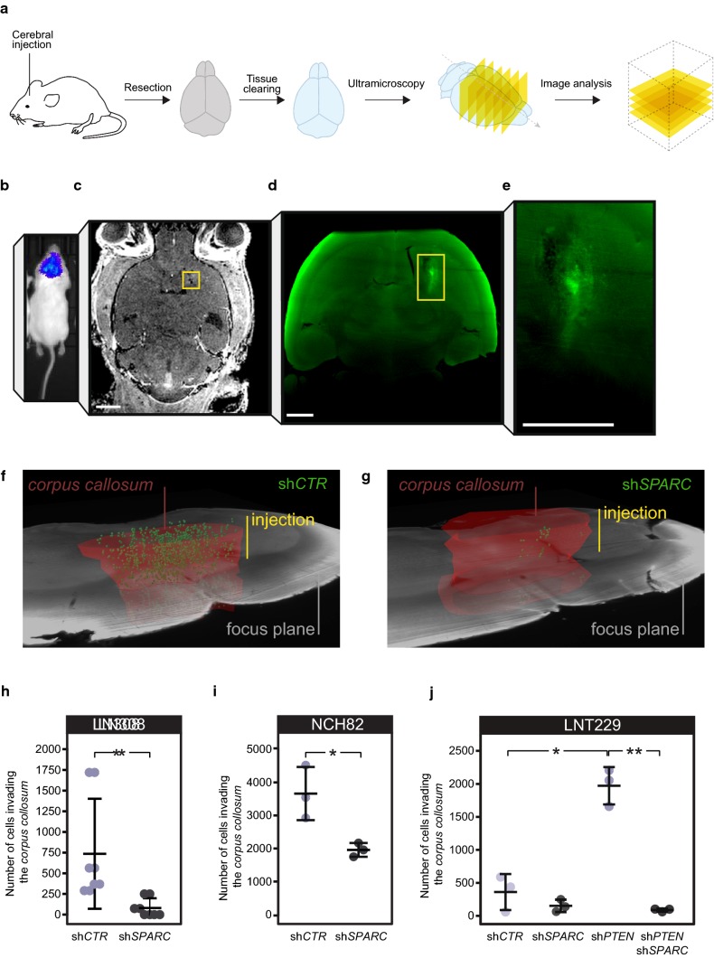
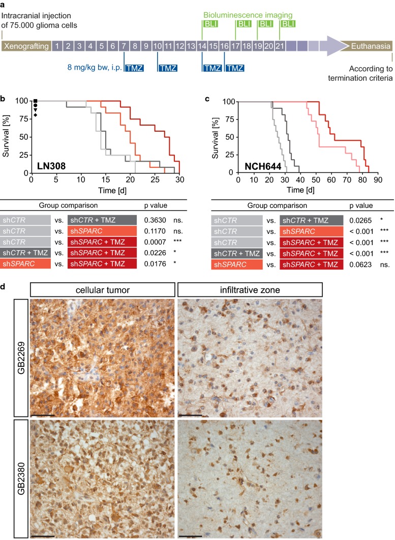
Glioblastomas strongly invade the brain by infiltrating into the white matter along myelinated nerve fiber tracts even though the myelin protein Nogo-A prevents cell migration by activating inhibitory RhoA signaling. The mechanisms behind this long-known phenomenon remained elusive so far, precluding a targeted therapeutic intervention. This study demonstrates that the prevalent activation of AKT in gliomas increases the ER protein-folding capacity and enables tumor cells to utilize a side effect of RhoA activation: the perturbation of the IRE1α-mediated decay of SPARC mRNA. Once translation is initiated, glioblastoma cells rapidly secrete SPARC to block Nogo-A from inhibiting migration via RhoA. By advanced ultramicroscopy for studying single-cell invasion in whole, undissected mouse brains, we show that gliomas require SPARC for invading into white matter structures. SPARC depletion reduces tumor dissemination that significantly prolongs survival and improves response to cytostatic therapy. Our finding of a novel RhoA-IRE1 axis provides a druggable target for interfering with SPARC production and underscores its therapeutic value.

Keywords: AKT; ENTPD5; Glioblastoma; IRE1α; Invasion; Nogo-A; Post-transcriptional regulation; RhoA; SPARC; White matter.


Conflict of interest statement

GR has received research Grants from Roche and Merck Serono.

Figures

Fig. 1
Fig. 1
Glioblastoma cells respond to Nogo-A by activating S1PR2 and its downstream effector RhoA. a Nogo-A activates S1PR2 via its Δ20 domain. b S1PR2 and NgR1 levels in human brain and glioma cell lines (LN18, LNT229, LN308, LN443, LN446, and T98G) and in low passage patient-derived glioblastoma cells (NCH82, NCH89, NCH342, and NCH417). c, d13GTP levels in c LNT229 cells treated with 1 µM JTE-013 or d LN308 cells treated with blocking peptides mimicking ECL2 or ECL3 or a scrambled peptide (SCR). Error bars represent the SD, n = 3. Unpaired t-test, *p ≤ 0.05; **p ≤ 0.01; ***p ≤ 0.001; not significant = p > 0.05. e S1PR2 activation by Nogo-A induces RhoA. e, f RhoAGTP levels in LN308 cells. Control shRNA (shCTR); shRNA against S1PR2 (shS1PR2); shRNA against GNA13 encoding Gα13 (shGNA13). c, d, e, f Cells were seeded on Δ20-coated dishes and harvested for activity assays after 30 min (Gα13) or 1 h (RhoA). Nogo-A-Δ20 (Δ20); Nogo-A-ΔSCR (ΔSCR)
Fig. 2
Fig. 2
Glioblastoma cells secrete the Nogo-A decoy SPARC upon RhoA activation. a, b, h Cells were grown on protein-coated surfaces for 16 h. a SPARC levels in secreted lysates and intracellular protein isolates from LN308 cells. b CLSM of LN308 cells. Scale bar: 20 µm. c, d RhoAGTP or SPARC levels in NCH343 cells grown at either low (2 × 104 cells/cm2) or high (10 × 104 cells/cm2) density. e, f, g IMAC using e TrxA-SPARC and human brain and liver lysates, f His-tagged Δ20 and EGFP-tagged SPARC with deletion of either the EGF-like motif (del_EC) or the Kazal-like motif (del_Kazal), or g RFP-tagged Δ20 min or Δ20 and SPARC-EGFP. The plot shows the propensity for intrinsic disorder within Nogo-A-Δ20. h SPARC levels in the presence of RFP-Δ20 min or a scrambled version. i Co-IP of Nogo-A, S1PR2, and SPARC using brain lysates from mice xenografted without glioblastoma cells (nx) or with LN308 glioblastoma cells expressing shCTR or shSPARC. Control shRNA (shCTR); shRNA against SPARC (shSPARC). f, g, h Nogo-A(Δ20) (Δ20); RFP-tagged minimal deleted Nogo-A(Δ20) (Δ20 min-RFP); scrambled Nogo-A(Δ20) (ΔSCR)
Fig. 3
Fig. 3
RhoA-induced deactivation of IRE1α initiates SPARC translation. a The canonical Rho-ROCK pathway is triggered by Nogo-A-mediated activation of S1PR2. b SPARC, MLC-2, and p-MLC-2S19 levels in LN308 cells. c, e SPARC levels in LN443 cells expressing RhoAG14V and treated with either 1 µM blebbistatin (Bleb) or 250 ng/ml latrunculin A (Lat-A) for 16 h. d p-MLC-2S19 levels in LN308 cells harvested after 1 h. Δ20/ΔSCR was pre-adsorbed with equimolar amounts of either TrxA-SPARC (SPARC) or TrxA-SPARC(del_Kazal) (del_Kazal). f Perturbation of IRE1α activity initiates SPARC translation. g, h SPARC levels in LN308 cells expressing control shRNA (shCTR) or shRNA against ERN1 (shERN1), which encodes IRE1α. (h) Gene expression analysis of ERN1 and SPARC. i SPARC levels in LN308 cells expressing either IRE1αS724A, IRE1αK907A or EGFP fused to an N-terminal signal peptide (SP-EGFP). j SPARC levels in LN443 cells expressing RhoAG14V and treated with 1 µM APY-29 for 16 h. k, l Total- and phospho-IRE1αS724 levels in LN308 cells exposed to Δ20 (k) or expressing RhoAG14V and treated with 1 µM Y-27632 (l). Error bars represent the SD, n = 3. Unpaired t test, *p ≤ 0.05; **p ≤ 0.01; ***p ≤ 0.001; not significant = p > 0.05. b, k Nogo-A-ΔSCR (ΔSCR); Nogo-A-Δ20 (Δ20)
Fig. 4
Fig. 4
Increased ENTPD5 expression due to high p-AKT levels allows for SPARC production. a, b ENTPD5 levels in a LN308 glioblastoma cells expressing either myc-tagged AKTS473A or PTEN; b LNT229 cells expressing either AKT1-myc, control shRNA (shCTR) or shRNA against PTEN (shPTEN). c ENTPD5 and SPARC levels in glioblastoma cells treated with MK-2206 for 16 h. d, e SPARC levels in d LN308 cells expressing ENTPD5E127A-myc or e LNT229 cells expressing ENTPD5-myc. c, d, e Cells were exposed to either Nogo-A-Δ20 (Δ20) or Nogo-A-ΔSCR (ΔSCR) for 16 h. f SPARC levels in glioblastoma cells expressing PTEN-myc were treated with 1 µM 4µ8C. g The RhoA-activated perturbation of IRE1α-regulated mRNA decay (RAPID) pathway
Fig. 5
Fig. 5
Glioblastoma cells require SPARC to migrate on myelinated structures in vitro. a, b Migration of a LNT229 or LN308 cells in the presence of increasing myelin concentrations or of b LNT229 cells in the presence of 20 µg/cm2 myelin blocked with increasing concentrations of α-Nogo-A antibody. c, d, e Real-time cell analysis. Transwells were coated with 5 µg/cm2 Nogo-A-Δ20 (Δ20). LN308 cell migration in the presence of c JTE-013 or d APY-29. e NCH82 cell migration in the presence of equimolar amounts of SPARC and/or 1 µM MK-2206. f LNT229 cell migration in the presence of increasing concentrations of either SPARC or SPARC-del_Kazal (del_Kazal). a, b, c, d, e, f Unpaired t-test, error bars represent the SD, *p ≤ 0.05; **p ≤ 0.01; ***p ≤ 0.001; ns. = p > 0.05
Fig. 6
Fig. 6
Glioblastoma cells require SPARC to invade white matter in vivo. a SPIM workflow for ultramicroscopic analysis of glioma cell invasion. b, c, d, e Increased resolution from b BLI, c MRT with injection site (yellow square); scale bar: 2 mm, d, e UM; scale bar: 100 μm. f, g UM analysis of brains xenografted with LN308EGFP−2A−FLuc cells. The focus plane is indicated in gray, and the injection site on the lateral side to the corpus callosum (red) is indicated in yellow. h, i, j Quantification of EGFP-labelled h LN308 cells, i NCH82 cells, and j LNT229 cells that invaded the corpus callosum. Wilcoxon rank-sum test, error bars represent the SD, *p ≤ 0.05; **p ≤ 0.01; ***p ≤ 0.001; ns. = p > 0.05. Control shRNA (shCTR), shRNA against SPARC (shSPARC), shRNA against PTEN (shPTEN)
Fig. 7
Fig. 7
Glioblastoma cells require SPARC for infiltrative growth. a Treatment scheme of xenografted NSG mice. b, c Kaplan–Meier analysis of NSG mice xenografted with LN308 or NCH644 cells expressing EGFP-2A-FLuc and treated with temozolomide (TMZ) or DMSO. Control shRNA (shCTR), shRNA against SPARC (shSPARC). Mice that died due to a non-tumor-related cause were censored by a symbol: shCTR (circle), shCTR + TMZ (square), shSPARC (triangle), shSPARC + TMZ (rhombus). d Immunohistochemical staining for SPARC in tissue samples from two representative IDH-wildtype glioblastomas, with (GB2380) or without PTEN mutation (GB2269). Shown are images of cellular tumor areas (left column) and the respective infiltration zone (right column). Brown: specific immunoreactivity. Blue: counterstaining with hemalum. Scale bars: 50 µM

Similar articles

Cited by

References

    1. Bady P, Diserens AC, Castella V, Kalt S, Heinimann K, Hamou MF, et al. DNA fingerprinting of glioma cell lines and considerations on similarity measurements. Neuro Oncol. 2012;14:701–711. doi: 10.1093/neuonc/nos072. - DOI - PMC - PubMed
    1. Breckwoldt MO, Bode J, Kurz FT, Hoffmann A, Ochs K, Ott M, et al. Correlated magnetic resonance imaging and ultramicroscopy (MR-UM) is a tool kit to assess the dynamics of glioma angiogenesis. Elife. 2016;5:e11712. doi: 10.7554/eLife.11712. - DOI - PMC - PubMed
    1. Caroni P, Schwab ME. Antibody against myelin-associated inhibitor of neurite growth neutralizes nonpermissive substrate properties of CNS white matter. Neuron. 1988;1:85–96. doi: 10.1016/0896-6273(88)90212-7. - DOI - PubMed
    1. Chalhoub N, Baker SJ. PTEN and the PI3-kinase pathway in cancer. Ann Rev Pathol. 2009;4:127–150. doi: 10.1146/annurev.pathol.4.110807.092311. - DOI - PMC - PubMed
    1. Chlenski A, Cohn SL. Modulation of matrix remodeling by SPARC in neoplastic progression. Semin Cell Dev Biol. 2010;21:55–65. doi: 10.1016/j.semcdb.2009.11.018. - DOI - PubMed

Publication types

MeSH terms