Observational Study
. 2020 Feb;38(2):417-424.
doi: 10.1007/s00345-019-02793-9. Epub 2019 May 6.

Organ-sparing surgery of penile cancer: higher rate of local recurrence yet no impact on overall survival

Affiliations
Observational Study

Organ-sparing surgery of penile cancer: higher rate of local recurrence yet no impact on overall survival

Andrea Katharina Lindner et al. World J Urol. 2020 Feb.

Abstract

Purpose: To report on the oncological outcome of organ-sparing surgery (OSS) compared to (total or partial) penectomy regarding recurrence patterns and survival in squamous cell carcinoma (SCC) of the penis.

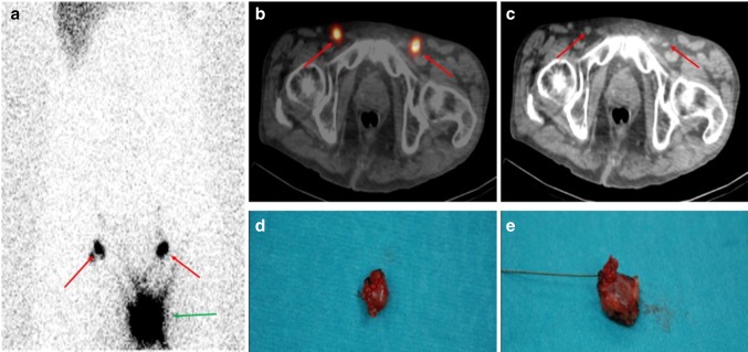
Methods: This was a retrospective study of all patients with penile SCC and eligible follow-up data of at least 2 years at our institution. Patients with tumors staged ≥ pT1G2 underwent invasive lymph node (LN) staging by dynamic sentinel-node biopsy or modified inguinal lymphadenectomy. Radical inguinal lymphadenectomy was performed when LNs were palpable at diagnosis and in those with a positive LN status after invasive nodal staging. Follow-up visits were assessed, and local, regional and distant recurrences were defined and analyzed.

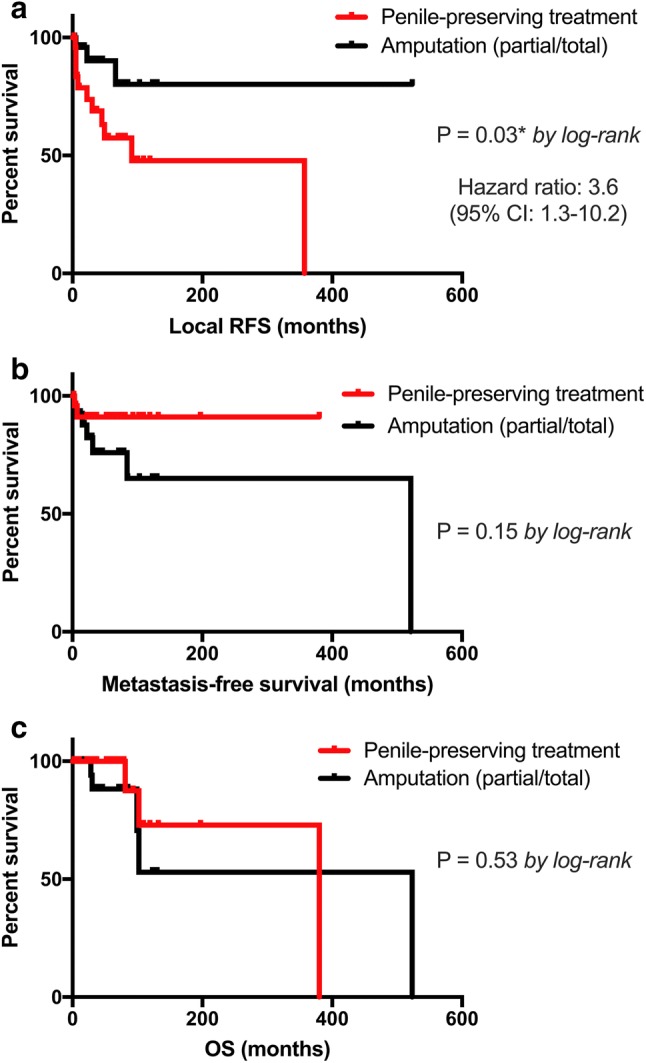
Results: 55 patients were identified with a mean follow-up of 63.7 months. Surgical management was OSS in 26 patients (47.2%) and partial or total penectomy in 29 cases (52.8%). Histopathological staging was: pTis (12.7%), pTa (16.3%), pT1a (18.2%), pT1b (5.5%), pT2 (29.1%) and pT3 (18.2%), respectively. Patients in the penectomy group were significantly older (mean 68 vs. 62 years; p = 0.026) with a higher rate of advanced tumor stage (≥ pT2: 44.8% vs. 11.5%; p = 0.002). The local recurrence rate was 42.3% (n = 11) following OSS compared to 10.3% (n = 3) after penectomy (p = 0.007). Kaplan-Meier curves showed no significant differences between the two groups regarding metastasis-free and overall survival.

Conclusions: OSS is associated with a higher local recurrence rate compared to penectomy, yet it has no negative impact on overall and metastasis-free survival.

Keywords: Follow-up; Organ-preserving surgery; Penile neoplasm; Recurrence; Squamous cell; Survival.


Conflict of interest statement

The authors declare that there is no conflict of interest regarding the publication of this paper.

Figures

Fig. 1
Fig. 1
Sentinel lymph node biopsy (SLNB) guided by lymphoscintigraphy [39]. In addition to early dynamic imaging (not displayed) following intradermal injection of 40 MBq Technetium-99 m-labelled nanocolloidal albumin (99mTc-Nanocoll®) peritumorally, planar static imaging 60 min post-injection of the pelvis is performed, including combined single-photon-emission tomography with low-dose computed tomography (SPECT/CT) acquisition. On planar image 60 min p.i. apart from the injection site (a green arrow), intense focal tracer uptake is visualized in the right and left pelvic area (a red arrows). On fused SPECT/CT images (b axial slice, red arrows), focal uptake is located in the inguinal region, that corresponded to non-enlarged lymph nodes on low-dose CT (c axial slice, red arrows), representing inguinal SLN. Both SLN were localized intra-operatively with a gamma-probe 1 day after tracer injection and could be surgically removed (d, e). Final histology confirmed pN0
Fig. 2
Fig. 2
Kaplan–Meier survival curves. a Local recurrence-free survival (RFS), b (lymphatic and hematogenous) metastasis-free survival and c overall survival (OS) in months according to the type of penile surgical approach (OSS vs. penile amputation). p values by log-rank test; *p < 0.05; **p < 0.01; ***p < 0.001

References

    1. Heller DS. Lesions and neoplasms of the penis: a review. J Low Genit Tract Dis. 2016;20(1):107–111. doi: 10.1097/LGT.0000000000000159. - DOI - PubMed
    1. Coelho RWP, Pinho JD, Moreno JS, Garbis DVEO, do Nascimento AMT, Larges JS, et al. Penile cancer in Maranhão, Northeast Brazil: the highest incidence globally? BMC Urol. 2018;18(1):50. doi: 10.1186/s12894-018-0365-0. - DOI - PMC - PubMed
    1. Barnholtz-Sloan JS, Maldonado JL, Pow-sang J, Guiliano AR. Incidence trends in primary malignant penile cancer. Urol Oncol Semin Orig Investig. 2007;25(5):361–367. doi: 10.1016/j.urolonc.2006.08.029. - DOI - PubMed
    1. Mosconi AM, Roila F, Gatta G, Theodore C. Cancer of the penis. Crit Rev Oncol Hematol. 2005;53(2):165–177. doi: 10.1016/j.critrevonc.2004.09.006. - DOI - PubMed
    1. Hakenberg OW, Compérat EM, Minhas S, Necchi A, Protzel C, Watkin N. EAU guidelines on penile cancer: 2014 update. Eur Urol. 2015;67(1):142–150. doi: 10.1016/j.eururo.2014.10.017. - DOI - PubMed

Publication types