. 2019 May 1;75(Pt 5):467-474.
doi: 10.1107/S2059798319005011. Epub 2019 Apr 30.

Visualizing membrane trafficking through the electron microscope: cryo-tomography of coat complexes

Affiliations

Visualizing membrane trafficking through the electron microscope: cryo-tomography of coat complexes

Evgenia A Markova et al. Acta Crystallogr D Struct Biol. .

Abstract

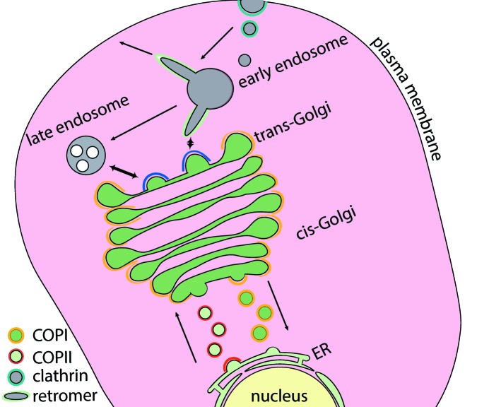
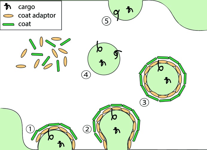
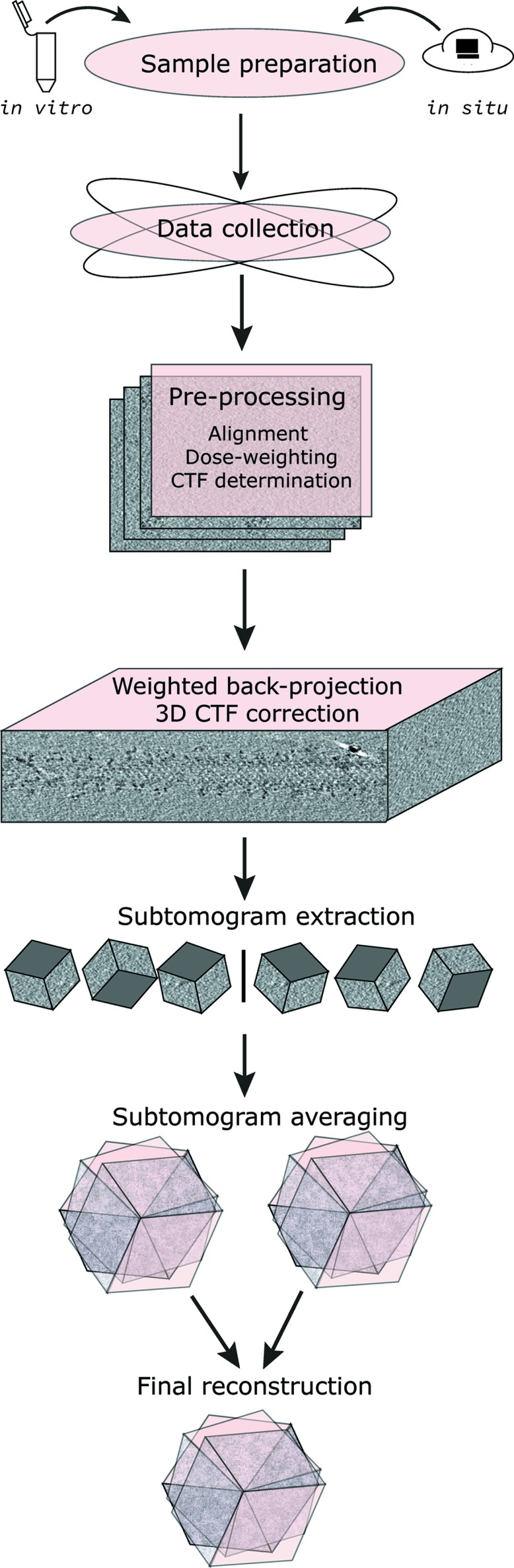
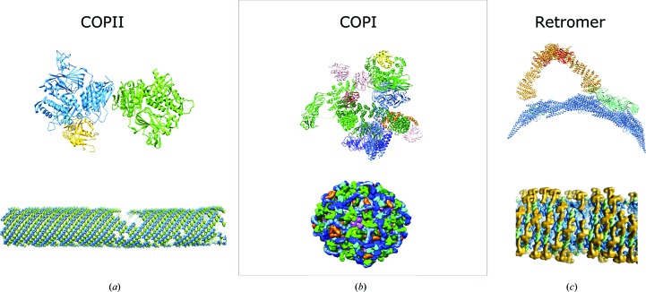
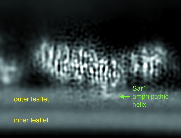
Coat proteins mediate vesicular transport between intracellular compartments, which is essential for the distribution of molecules within the eukaryotic cell. The global arrangement of coat proteins on the membrane is key to their function, and cryo-electron tomography and subtomogram averaging have been used to study membrane-bound coat proteins, providing crucial structural insight. This review outlines a workflow for the structural elucidation of coat proteins, incorporating recent developments in the collection and processing of cryo-electron tomography data. Recent work on coat protein I, coat protein II and retromer performed on in vitro reconstitutions or in situ is summarized. These studies have answered long-standing questions regarding the mechanisms of membrane binding, polymerization and assembly regulation of coat proteins.

Keywords: COPII; coat proteins; cryo-electron tomography; subtomogram averaging; three-dimensional reconstruction; vesicular transport.


Figures

Figure 1
Figure 1
The intracellular function of coat proteins. Coat proteins are responsible for the exchange of biomolecules between membrane-bound compartments and facilitate endocytosis and exocytosis. COPII vesicles transport cargo from the endoplasmic reticulum (ER) to the Golgi apparatus (red), while COPI vesicles are responsible for intra-Golgi transport and trafficking from the Golgi to the ER (yellow). Retromer enables the tubulation of the early endosome towards the trans-Golgi network (TGN) and the cell exterior (green). Clathrin coats endocytic vesicles, which are formed at the plasma membrane, and buds emerging from the trans-Golgi network (blue).
Figure 2
Figure 2
Vesicle budding and fusion. (1) Coat proteins are recruited to donor membranes, which have a characteristic lipid and protein composition and local curvature. Typically, a membrane-and-cargo-adapter-like layer and a coat-like membrane layer are assembled. Coat proteins and additional components concentrate cargo in the forming bud. (2) Membrane curvature increases and a vesicle neck forms. (3) Vesicle scission results in the release of the cargo-laden vesicle from the donor compartment. (4) Vesicle uncoating occurs, allowing subsequent fusion with the acceptor compartment. Coat proteins are released for recycling. (5) Fusion with the recipient compartment occurs and the release of cargo.
Figure 3
Figure 3
A cryo-electron tomography workflow used for the structural determination of COPII assembled on membranes.
Figure 4
Figure 4
In vitro reconstituted COPII assemblies on membranes. The addition of purified COPII components to giant unilamellar vesicles results in COPII membrane binding and the generation of a variety of morphologies, including beads-on-a-string (dashed line) and tubular (unbroken line) COPII assemblies. (a) COPII morphologies seen in a medium-magnification image of a cryo-grid. (b, c) Slices through binned 8× cryo-tomograms with 50 iterations of SIRT-like filtering applied, showing the regular lattice assembly of inner (b) and outer (c) coat components. Tomogram images are courtesy of Joshua Hutchings (unpublished work).
Figure 5
Figure 5
Structural insights gained from the study of coat proteins. The top panels represent fitted structural models of asymmetric units. The bottom panels show the global coat arrangement. (a) Inner COPII coat (Hutchings et al., 2018; PDB entry 6gni). Colour scheme: Sec23, blue; Sec24, green; Sar1, yellow. (b) COPI (Dodonova et al., 2015, 2017; PDB entry 5nzr). Colour scheme: Arf1, pink; γ-COP, light green; β-COP, dark green; ζ-COP, yellow; δ-COP, orange; β′-COP, light blue; α-COP, dark blue. (c) Retromer (Kovtun et al., 2018; PDB entry 6h7w). Colour scheme: Vps5, blue; Vps29, red; Vps35, yellow; Vps26, green. The images are reproduced with permission from AAAS and Springer Nature.
Figure 6
Figure 6
Association of the Sar1 GTPase with the ER membrane as seen in EMDB entry EMD-0044 (Hutchings et al., 2018 ▸). Sar1 inserts its N-­terminal helix into the membrane using a helical hook bent by 90°.

References

    1. Albert, S., Schaffer, M., Beck, F., Mosalaganti, S., Asano, S., Thomas, H. F., Plitzko, J. M., Beck, M., Baumeister, W. & Engel, B. D. (2017). Proc. Natl Acad. Sci. USA, 114, 13726–13731. - PMC - PubMed
    1. Antonny, B., Madden, D., Hamamoto, S., Orci, L. & Schekman, R. (2001). Nature Cell Biol. 3, 531–537. - PubMed
    1. Asano, S., Fukuda, Y., Beck, F., Aufderheide, A., Förster, F., Danev, R. & Baumeister, W. (2015). Science, 347, 439–442. - PubMed
    1. Bacia, K., Futai, E., Prinz, S., Meister, A., Daum, S., Glatte, D., Briggs, J. A. G. & Schekman, R. (2011). Sci. Rep. 1, 17. - PMC - PubMed
    1. Barlowe, C., Orci, L., Yeung, T., Hosobuchi, M., Hamamoto, S., Salama, N., Rexach, M. F., Ravazzola, M., Amherdt, M. & Schekman, R. (1994). Cell, 77, 895–907. - PubMed

MeSH terms

Substances