. 2019 Nov;70(5):1714-1731.
doi: 10.1002/hep.30700. Epub 2019 Jun 21.

Hippo Signaling Controls NLR Family Pyrin Domain Containing 3 Activation and Governs Immunoregulation of Mesenchymal Stem Cells in Mouse Liver Injury

Affiliations

Hippo Signaling Controls NLR Family Pyrin Domain Containing 3 Activation and Governs Immunoregulation of Mesenchymal Stem Cells in Mouse Liver Injury

Changyong Li et al. Hepatology. 2019 Nov.

Abstract

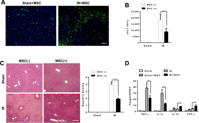
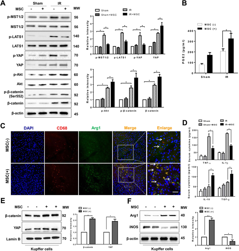
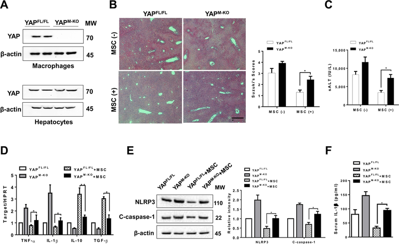
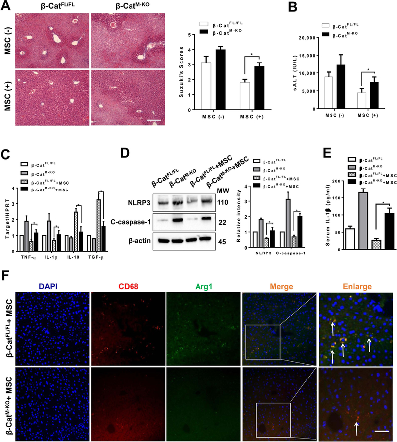
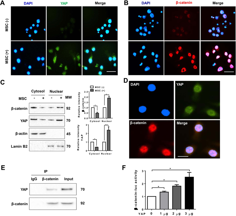
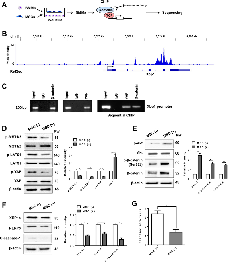
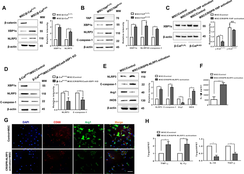
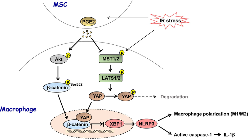
The Hippo pathway, an evolutionarily conserved protein kinase cascade, tightly regulates cell growth and survival. Activation of yes-associated protein (YAP), a downstream effector of the Hippo pathway, has been shown to modulate tissue inflammation. However, it remains unknown as to whether and how the Hippo-YAP signaling may control NLR family pyrin domain containing 3 (NLRP3) activation in mesenchymal stem cell (MSC)-mediated immune regulation during liver inflammation. In a mouse model of ischemia/reperfusion (IR)-induced liver sterile inflammatory injury, we found that adoptive transfer of MSCs reduced hepatocellular damage, shifted macrophage polarization from M1 to M2 phenotype, and diminished inflammatory mediators. MSC treatment reduced mammalian Ste20-like kinase 1/2 and large tumor suppressor 1 phosphorylation but augmented YAP and β-catenin expression with increased prostaglandin E2 production in ischemic livers. However, disruption of myeloid YAP or β-catenin in MSC-transferred mice exacerbated IR-triggered liver inflammation, enhanced NLRP3/caspase-1 activity, and reduced M2 macrophage phenotype. Using MSC/macrophage coculture system, we found that MSCs increased macrophage YAP and β-catenin nuclear translocation. Importantly, YAP and β-catenin colocalize in the nucleus while YAP interacts with β-catenin and regulates its target gene X-box binding protein 1 (XBP1), leading to reduced NLRP3/caspase-1 activity after coculture. Moreover, macrophage YAP or β-catenin deficiency augmented XBP1/NLRP3 while XBP1 deletion diminished NLRP3/caspase-1 activity. Increasing NLRP3 expression reduced M2 macrophage arginase1 but augmented M1 macrophage inducible nitric oxide synthase expression accompanied by increased interleukin-1β release. Conclusion: MSCs promote macrophage Hippo pathway, which in turn controls NLRP3 activation through a direct interaction between YAP and β-catenin and regulates XBP1-mediated NLRP3 activation, leading to reprograming macrophage polarization toward an anti-inflammatory M2 phenotype. Moreover, YAP functions as a transcriptional coactivator of β-catenin in MSC-mediated immune regulation. Our findings suggest a therapeutic target in MSC-mediated immunotherapy of liver sterile inflammatory injury.


Figures

Figure 1
Figure 1. Adoptive transfer of MSCs attenuates IR-induced liver injury and inhibits proinflammatory mediator program.
Mice were subjected to 90min of partial liver warm ischemia, followed by 6h of reperfusion. Some animals were injected via tail vein with MSCs (1×10⁶) 24h prior to ischemia insult. (A) MSCs were labeled with 5-CMFDA to track the distribution of MSCs in ischemic livers. Representative immunofluorescence staining for the MSCs labeled with 5-CMFDA (green) localized in IR-stressed livers after MSC treatment (n=3–4 mice/group). DAPI was used to visualize nuclei (blue). Scale bars, 20μm. (B) Hepatocellular function was evaluated by sALT levels (IU/L) (n=4–6 samples/group). (C) Representative histological staining (H&E) of ischemic liver tissue (n=4–6 mice/group) and Suzuki’s histological score. Scale bars, 100μm. (D) qRT-PCR-assisted detection of TNF-α, IL-1β, IL-10, and TGF-β in ischemic livers (n=3–4 samples/group). Data were normalized to HPRT gene expression. All data represent the mean±SD. *p<0.05, **p<0.01.
Figure 2
Figure 2. MSCs regulate Hippo signaling/β-catenin activation and control macrophage polarization in IR-stressed livers.
(A) Immunoblot-assisted analysis and relative density ratio of p-MST½, MST½, p-LATS1, LATS1, p-YAP, YAP, p-Akt, Akt, p-β-catenin, and β-catenin in IR-stressed livers with or without MSC treatment. Representative of three experiments. (B) ELISA analysis of PGE2 levels in animal serum (n=3–4 samples/group). (C) Representative immunofluorescence staining for the macrophage marker CD68 (red) and arginase-1 (Arg1, green) co-localization in IR-stressed livers (n=3–4 mice/group). DAPI was used to visualize nuclei (blue). Arrow indicated CD68 and Arg1 double positive macrophages (yellow). Scale bars, 20μm. (D) ELISA analysis of TNF-α, IL-1β, IL-10, and TGF-β levels in animal serum (n=3–4 samples/group). Immunoblot-assisted analysis and relative density ratio of nuclear YAP and β-catenin (E), and Arg1 and iNOS (F) in liver Kupffer cells. Representative of three experiments. All data represent the mean±SD. *p<0.05, **p<0.01.
Figure 3
Figure 3. Myeloid YAP deficiency in MSC-treated livers aggravates IR-induced hepatocellular damage and promotes NLRP3 inflammasome-driven inflammatory response.
The YAPFL/FL and YAPM-KO mice were subjected to 90min of partial liver warm ischemia, followed by 6h of reperfusion. Some animals were injected via tail vein with MSCs (1×10⁶) 24h prior to ischemia. (A) The YAP expression was detected in hepatocytes and liver macrophages (Kupffer cells) by Western blot assay. Representative of three experiments. (B) Representative histological staining (H&E) of ischemic liver tissue (n=4–6 mice/group) and Suzuki’s histological score. Scale bars, 100μm. (C) Hepatocellular function was evaluated by sALT levels (IU/L) (n=4–6 samples/group). (D) qRT-PCR-assisted detection of TNF-α, IL-1β, IL-10, and TGF-β in ischemic livers (n=3–4 samples/group). Data were normalized to HPRT gene expression. (E) Immunoblot-assisted analysis and relative density ratio of NLRP3 and cleaved caspase-1 in ischemic livers. Representative of three experiments. (F) ELISA analysis of IL-1β levels in animal serum (n=3–4 samples/group). All data represent the mean±SD. *p<0.05, **p<0.01.
Figure 4
Figure 4. Disruption of myeloid β-catenin in MSC-treated livers activates NLRP3 and diminishes M2 macrophage polarization in IR-stressed livers.
The β-cateninFL/FL and β-cateninM-KO mice were subjected to 90min of partial liver warm ischemia, followed by 6h of reperfusion. Some animals were injected via tail vein with MSCs (1×10⁶) 24h prior to ischemia. (A) Representative histological staining (H&E) of ischemic liver tissue (n=4–6 mice/group) and Suzuki’s histological score. Scale bars, 100μm. (B) Hepatocellular function was evaluated by sALT levels (IU/L) (n=4–6 samples/group). (C) qRT-PCR-assisted detection of TNF-α, IL-1β, IL-10, and TGF-β in ischemic livers (n=3–4 samples/group). Data were normalized to HPRT gene expression. (D) Immunoblot-assisted analysis and relative density ratio of NLRP3 and cleaved caspase-1 in ischemic livers. Representative of three experiments. (E) ELISA analysis of IL-1β levels in animal serum (n=3–4 samples/group). (F) Representative immunofluorescence staining for the macrophage marker CD68 (red) and arginase-1 (Arg1, green) co-localization in IR-stressed livers (n=3–4 mice/group). DAPI was used to visualize nuclei (blue). Arrow indicated CD68 and Arg1 double positive macrophages (yellow). Scale bars, 20 μm. All data represent the mean±SD. *p<0.05.
Figure 5
Figure 5. YAP interacts with β-catenin and regulates its transcription activity in MSC-mediated immune regulation.
Bone marrow-derived macrophages (BMMs, 1×10⁶) were co-cultured with MSCs (2×10⁵) for 24h followed by LPS (100 ng/ml) stimulation. (A) and (B) Immunofluorescence staining of nuclear YAP (green) and β-catenin (red) in macrophages after co-culture with or without MSCs. DAPI was used to visualize nuclei (blue). Scale bars, 20μm. (C) Immunoblot-assisted analysis of cytosol and nuclear YAP and β-catenin in macrophages after co-culture with or without MSCs. Representative of three experiments. (D) Immunofluorescence staining for macrophage YAP (green) and β-catenin (red) co-localization in the nucleus after co-culture with MSCs. DAPI was used to visualize nuclei (blue). Scale bars, 10μm. (E) Immunoprecipitation analysis of YAP and β-catenin in macrophages after co-culture with MSCs. Representative of three experiments. (F) BMMs were co-transfected with 1µg β-catenin-luc and CRISPR YAP activation vectors. The luciferase activity was measured after 48h (n=3–4 samples/group). Data represent the mean±SD. *p<0.05.
Figure 6
Figure 6. The YAP-β-catenin signaling targets XBP1 and inhibits NLRP3-driven inflammatory response in MSC-mediated immune regulation.
(A) Experimental design of β-catenin ChIP-seq analysis. BMMs were collected and fixed after co-culture with MSCs. Following chromatin shearing and β-catenin antibody selection, the precipitated DNA fragments bound by β-catenin-containing protein complexes were used for sequencing. (B) Localization of β-catenin-binding sites on the mouse xbp1 gene. The five exons, four introns, 3’ UTR, 5’ UTR and TSS of the mouse xbp1 gene on chromosome 11 are shown. (C) ChIP-PCR analysis of YAP and β-catenin binding to the Xbp1 promoter. Protein-bound chromatin was prepared from BMMs and immunoprecipitated with YAP or β-catenin antibodies. For sequential ChIP, the protein-bound chromatin was first immunoprecipitated with the β-catenin antibody followed by elution with a second immunoprecipitation using YAP antibody, and then the immunoprecipitated DNA was analyzed by PCR. The normal IgG was used as a negative control. (D) (E) (F) Immunoblot-assisted analysis and relative density ratio of p-MST½, MST½, p-LATS1, LATS1, p-YAP, YAP, p-Akt, Akt, p-β-catenin, β-catenin, XBP1s, NLRP3, and cleaved caspase-1 in macrophages after co-culture with or without MSCs. Representative of three experiments. (G) Caspase-1 activity (U) in macrophages after co-culture (n=3–4 samples/group). All data represent the mean±SD. *p<0.05, **p<0.01.
Figure 7
Figure 7. YAP is crucial to mediate β-catenin activity and reprograms NLRP3-dependent macrophage polarization in MSC-mediated immune regulation.
(A) (B) BMMs were isolated from β-cateninFL/FL, β-cateninM-KO, YAPFL/FL, YAPM-KO mice and then co-cultured with MSCs followed by LPS stimulation (n=3–4 samples/group). Immunoblot-assisted analysis and relative density ratio of macrophage β-catenin, YAP, XBP1s, and NLRP3, and cleaved caspase-1. Representative of three experiments. (C) BMMs were isolated from β-cateninFL/FL and β-cateninM-KO mice and transfected with CRISPR-mediated YAP activation or control vector, and then co-cultured with MSCs followed by LPS stimulation. Immunoblot-assisted analysis and relative density ratio of macrophage XBP1s. Representative of three experiments. (D) BMMs were isolated from β-cateninM-KO mice and transfected with CRISPR/Cas9-mediated XBP1 KO or control vector and then co-cultured with MSCs followed by LPS stimulation. Immunoblot-assisted analysis and relative density ratio of macrophage XBP1s, NLRP3, and cleaved caspase-1. Representative of three experiments. Fig. 7E-H, BMMs were isolated from β-cateninFL/FL mice and transfected with CRISPR-mediated NLRP3 activation or control vector were co-cultured with MSCs followed by LPS stimulation. (E) Immunoblot-assisted analysis and relative density ratio of macrophage NLRP3, cleaved caspase-1, Arg1, and iNOS. Representative of three experiments. (F) ELISA analysis of IL-1β levels in animal serum (n=3–4 samples/group). (G) Representative immunofluorescence staining for the macrophage marker CD68 (red) and arginase-1 (Arg1, green) co-localization in BMMs. DAPI was used to visualize nuclei (blue). Scale bars, 20μm. (H) qRT-PCR-assisted detection of TNF-α, IL-1β, IL-10, and TGF-β in macrophages (n=3–4 samples/group). Data were normalized to HPRT gene expression. All data represent the mean±SD. *p<0.05, **p<0.01.
Figure 8
Figure 8. Schematic illustration how Hippo signaling may control NLRP3 activation in MSC-mediated immune regulation.
IR stress increases MSC-mediated PGE2 section, which in turn activates macrophage Akt and phosphorylates β-catenin at Ser552 leading to translocation of β-catenin into nucleus. Notably, MSCs regulate macrophage Hippo-YAP pathway by depressing MST½ and LATS1 phosphorylation, and increasing YAP translocation from cytoplasm to nucleus where YAP co-localizes and interacts with nuclear β-catenin, which in turn regulates their target gene XBP1 leading to reduced NLRP3/caspase-1 activity and IL-1β release, and augmented M2 macrophage phenotype in IR-triggered liver inflammation.

References

    1. Dimarino AM, Caplan AI, Bonfield TL. Mesenchymal stem cells in tissue repair. Front Immunol 2013;4:201. - PMC - PubMed
    1. Wang LT, Ting CH, Yen ML, Liu KJ, Sytwu HK, Wu KK, Yen BL. Human mesenchymal stem cells (MSCs) for treatment towards immune- and inflammation-mediated diseases: review of current clinical trials. J Biomed Sci 2016;23:76. - PMC - PubMed
    1. Karp JM, Leng Teo GS. Mesenchymal stem cell homing: the devil is in the details. Cell Stem Cell 2009;4:206–216. - PubMed
    1. Zhao B, Tumaneng K, Guan KL. The Hippo pathway in organ size control, tissue regeneration and stem cell self-renewal. Nat Cell Biol 2011;13:877–883. - PMC - PubMed
    1. Saucedo LJ, Edgar BA. Filling out the Hippo pathway. Nat Rev Mol Cell Biol 2007;8:613–621. - PubMed

Publication types

MeSH terms

Substances