. 2019 Sep 6;21(9):1131-1140.
doi: 10.1093/neuonc/noz079.

Oncolytic HSV therapy increases trametinib access to brain tumors and sensitizes them in vivo

Affiliations

Oncolytic HSV therapy increases trametinib access to brain tumors and sensitizes them in vivo

Ji Young Yoo et al. Neuro Oncol. .

Abstract

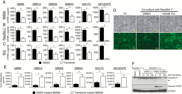
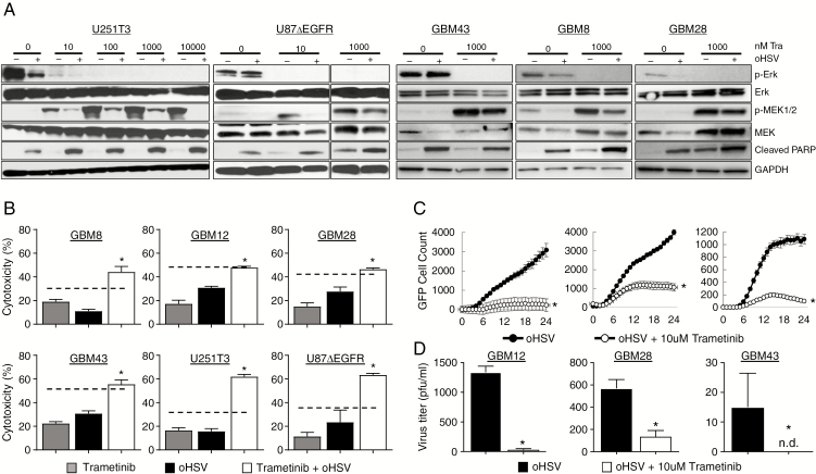
Background: Hyperactivation of the RAS-RAF-MEK-ERK signaling pathway is exploited by glioma cells to promote their growth and evade apoptosis. MEK activation in tumor cells can increase replication of ICP34.5-deleted herpes simplex virus type 1 (HSV-1), but paradoxically its activation in tumor-associated macrophages promotes a pro-inflammatory signaling that can inhibit virus replication and propagation. Here we investigated the effect of blocking MEK signaling in conjunction with oncolytic HSV-1 (oHSV) for brain tumors.

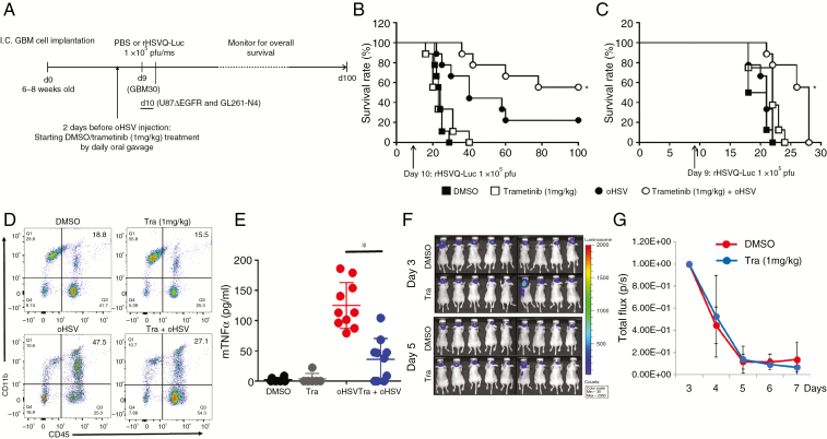
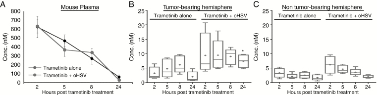
Methods: Infected glioma cells co-cultured with microglia or macrophages treated with or without trametinib were used to test trametinib effect on macrophages/microglia. Enzyme-linked immunosorbent assay, western blotting, and flow cytometry were utilized to evaluate the effect of the combination therapy. Pharmacokinetic (PK) analysis of mouse plasma and brain tissue was used to evaluate trametinib delivery to the CNS. Intracranial human and mouse glioma-bearing immune deficient and immune competent mice were used to evaluate the antitumor efficacy.

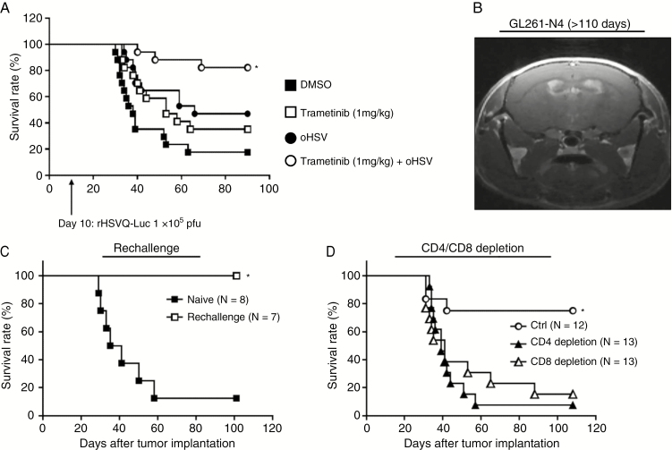
Result: Oncolytic HSV treatment rescued trametinib-mediated feedback reactivation of the mitogen-activated protein kinase signaling pathway in glioma. In vivo, PK analysis revealed enhanced blood-brain barrier penetration of trametinib after oHSV treatment. Treatment by trametinib, a MEK kinase inhibitor, led to a significant reduction in microglia- and macrophage-derived tumor necrosis factor alpha (TNFα) secretion in response to oHSV treatment and increased survival of glioma-bearing mice. Despite the reduced TNFα production observed in vivo, the combination treatment activated CD8+ T-cell mediated immunity and increased survival in a glioma-bearing immune-competent mouse model.

Conclusion: This study provides a rationale for combining oHSV with trametinib for the treatment of brain tumors.

Keywords: RAS-RAF-MEK-ERK signaling; glioblastoma (GBM); oncolytic herpes simplex virus-1 (oHSV); trametinib; tumor necrosis factor α (TNFα).


Figures

Fig. 1
Fig. 1
Trametinib treatment modulates macrophage and microglia anti-viral responses. (A‒C) TNFα release from BMDM (A), Raw264.7 (B), and BV2 (C) pretreated with or without trametinib (100 nM for 1 h), and then overlaid on the indicated primary GBM or glioma cell lines treated with saline or rHSVQ (MOI = 0.1), n = 3/group. (D‒E) Impact of trametinib on macrophage mediated virus clearance. (D) Fluorescent microscopy images of GFP-positive U251T3 human glioma cells, infected with rHSVQ (MOI = 0.01) for 1 h. After thorough washing to remove unbound virus particles, glioma cells were overlaid with DMSO or trametinib pretreated macrophage for 24 hours. Data shown are bright field and fluorescence microscopic images (bottom) of GFP-positive oHSV-infected cells 48 hours post infection. All bars are 100 μm for ×10 magnification images. (E) Quantification of virus yield from patient derived primary GBM and glioma cell lines infected with oHSV alone or co-cultured with primary BMDM cells. Briefly both cells and media were harvested 48 hours after overlay with DMSO or trametinib treated BMDM cells and viral titers were determined by standard plaque forming assay. Data shown are mean virus titer ±SD. *P < 0.05; n = 3/group. (F) Western blot analysis of co-cultures of U251T3 glioma cells with Raw264.7 macrophage cells pretreated with trametinib. U251T3 glioma cells were treated with/without rHSVQ (MOI = 0.5) for 1 h and then overlaid with macrophages pretreated with DMSO or trametinib overnight. Twenty-four hours post overlay, cells were harvested and cell lysates were probed with antibodies against caspase-8 and cleaved PARP. Βeta-tubulin was used as a loading control. Caspase-8 antibody can detect both total and cleaved forms of caspase-8. Arrow head indicates cleaved active form of caspase-8.
Fig. 2
Fig. 2
The combination effect of trametinib and oHSV on glioma cells. (A) Western blot analysis of the indicated cells treated with rHSVQ in the presence or absence of trametinib. The indicated patient derived primary GBM and glioma cells were infected with 0.1 MOI of rHSVQ. One hour post oHSV infection, unbound virus was removed and treated with the indicated concentration of trametinib (0.1‒10 μM). Sixteen hours post treatment, cells were harvested and cell lysates were probed with antibodies against pERK, pMEK1/2, total MEK1/2, total ERK, and cleaved PARP. Glyceraldehyde 3-phosphate dehydrogenase was used as a loading control. (B) Effects of trametinib and oHSV on glioma cell killing. The indicated cells were infected with oHSV or PBS for 1 hour, followed by treatment with trametinib (10 μM) or DMSO. Seventy-two hours later, cell viability was measured via standard MTT assay. Data shown are mean % of cell death ±SD and analyzed by the paired Student’s t-test (n = 3/group). Dotted line indicates the predicted additive cell killing. (C) Comparison of virus spread in cultures of the indicated glioma cells treated with rHSVQ in the presence or absence of trametinib over time. The indicated glioma cells were infected with rHSVQ and GFP expression was monitored every 2 hours for 24 hours utilizing the Cytation 5 live imaging system. GFP object count was quantified and graphed as an average of 4 wells per treatment group. Data shown are average counts of GFP positive cells ±SD over time. (D) Both cells and media were collected 48 hours post virus infection and virus titers were measured by standard plaque forming assay. Data shown are median titers ±SD (n = 3/group). (N.D. is not detectable).
Fig. 3
Fig. 3
Impact of trametinib on antitumor efficacy of oHSV in vivo. (A) Schematic representation of animal survival study. (B‒C) Kaplan‒Meier survival curve of mice bearing intracranial tumors treated with DMSO or trametinib and treated with phosphate buffered saline (PBS) or oHSV. Briefly, athymic nude mice bearing intracranial human glioma cells (B‒C) were treated with either DMSO or trametinib (1 mg/kg) by daily oral gavage. Two days after the initiation of gavage (10 days post tumor implantation for U87ΔEGFR) and 9 days post tumor implantation for GBM30), mice were treated with PBS or rHSVQ-Luc (1 × 10⁵ pfu) by intratumoral injection (n = 10/each groups for B‒C). Data shown are Kaplan‒Meier survival curves of mice treated with oHSV and/or trametinib. *P < 0.05 compared with mice receiving single treatment of oHSV. The arrows indicate the time of treatment with oHSV. (D‒G) Intracranial glioma (U87ΔEGFR) bearing mice were treated with DMSO or trametinib (1 mg/kg) by daily oral gavage, starting from day 8 post tumor cell implantation. Ten days post tumor cell implantation, mice were injected with rHSVQ-Luc by direct intratumoral injection. (D) Three days post virus injection, tumor-bearing hemispheres were analyzed for CD11bhigh/CD45+ monocyte derived macrophage infiltration and activation by flow cytometry. Data shown are a representative scatter plot showing CD11bhigh/CD45+ monocyte derived macrophage infiltration in vivo. (E) TNFα levels in brain hemispheres of mice treated with oHSV and trametinib. Twenty-four hours post oHSV injection, tumor-bearing brain hemispheres were homogenized and the supernatants were analyzed for murine TNFα by ELISA (DMSO and trametinib, n = 6; oHSV and trametinib plus oHSV, n = 10). Combination treatment with trametinib and oHSV significantly suppressed murine TNFα compared with oHSV alone. (F‒G) Trametinib effect on virus replication over time. (F) Representative bioluminescence images of mice treated with rHSVQ-Luc showing virus-encoded luciferase activity in mice treated with trametinib or DMSO at the indicated days post virus injection. (G) Quantification of images shown in F, Data shown are mean ±SD of quantification of total flux in each mouse, (n = 9/group).
Fig. 4
Fig. 4
Impact of oHSV therapy on trametinib brain tumor penetration in vivo. Nude mice bearing intracranial U87ΔEGFR human gliomas were treated as described above. Three days post virus injection, mice were sacrificed at the indicated time following the fifth trametinib treatment and plasma and both brain hemispheres were harvested (n = 5 mice/group). Trametinib concentration in plasma and brain was quantified by LC-MS/MS assay. Concentration-time plot of main PK trametinib in mouse plasma (A), tumor-bearing brain hemisphere (B), and non-tumor-bearing brain hemisphere (C). *P < 0.05 compared with mice treated with single treatment with oHSV.
Fig. 5
Fig. 5
Combination treatment with trametinib and oHSV enhances T-cell mediated antitumor efficacy. (A) C57BL/6 mice bearing syngeneic murine glioma GL261-N4 cells were treated with either DMSO or trametinib (1 mg/kg) by daily oral gavage. Two days after initiation of gavage (10 days post tumor implantation), mice were treated with phosphate buffered saline or rHSVQ-Luc (1 × 10⁵ pfu) by intratumoral injection (n = 17/each groups). Data shown are Kaplan‒Meier survival curves of mice treated with oHSV and/or trametinib. *P < 0.05 compared with mice receiving single treatment with oHSV. The arrows indicate the time of treatment of oHSV. (B) Representative MRI image of a C57BL/6 mouse treated with oHSV and trametinib in Fig. 5A 110 days after tumor cell implantation. (C) Mice treated with the combination trametinib and oHSV and surviving more than 100 days were rechallenged with a second intracranial GL261-N4 injection into the left hemisphere and compared with age matched naïve C57BL/6 mice (n = 7). Mice were monitored for up to 100 days and survival was analyzed by Kaplan–Meier. *P < 0.05. (D) Intracranial GL261-N4-bearing C57BL/6 mice were treated with trametinib and oHSV as described above as well as with isotype control, anti-CD4, or anti-CD8 depleting antibodies by intraperitoneal injection 2, 4, and 7 days post virus therapy (isotype control, n = 13; anti-CD4, n = 14; anti-CD8, n = 14). *P < 0.05 compared with CD4 or CD8 depletion.

Comment in

References

    1. Jo J, Wen PY. Antiangiogenic therapy of high-grade gliomas. Prog Neurol Surg. 2018;31:180–199. - PubMed
    1. Saha D, Wakimoto H, Rabkin SD. Oncolytic herpes simplex virus interactions with the host immune system. Curr Opin Virol. 2016;21:26–34. - PMC - PubMed
    1. Sanchala DS, Bhatt LK, Prabhavalkar KS. Oncolytic herpes simplex viral therapy: a stride toward selective targeting of cancer cells. Front Pharmacol. 2017;8:270. - PMC - PubMed
    1. Alvarez-Breckenridge C, Kaur B, Chiocca EA. Pharmacologic and chemical adjuvants in tumor virotherapy. Chem Rev. 2009;109(7):3125–3140. - PMC - PubMed
    1. Long GV, Stroyakovskiy D, Gogas H, et al. Dabrafenib and trametinib versus dabrafenib and placebo for Val600 BRAF-mutant melanoma: a multicentre, double-blind, phase 3 randomised controlled trial. Lancet. 2015;386(9992):444–451. - PubMed

Publication types

MeSH terms