. 2019 Aug;46(8):743-751.
doi: 10.1111/1440-1681.13101. Epub 2019 May 23.

OBG-like ATPase 1 inhibition attenuates angiotensin II-induced hypertrophic response in human ventricular myocytes via GSK-3beta/beta-catenin signalling

Affiliations

OBG-like ATPase 1 inhibition attenuates angiotensin II-induced hypertrophic response in human ventricular myocytes via GSK-3beta/beta-catenin signalling

Gayathri Narasimhan et al. Clin Exp Pharmacol Physiol. 2019 Aug.

Abstract

Obg-like ATPase 1 (OLA1) that possesses both GTP and ATP hydrolyzing activities has been shown to be involved in translational regulation of cancer cell growth and survival. Also, GSK3β signalling has been implicated in cardiac development and disease. However, the role of OLA1 in pathological cardiac hypertrophy is unknown. We sought to understand the mechanism by which OLA1 regulates GSK3β-β-Catenin signalling and its functional significance in angiotensin-II (ANG II)-induced cardiac hypertrophic response. OLA1 function and its endogenous interaction with GSK3β/β-catenin signalling in cultured human ventricular cardiomyocytes (AC16 cells) and mouse hearts (in vivo) was evaluated with/without ANG II-stimulated hypertrophic response. ANG II administration in mice increases myocardial OLA1 protein expression with a corresponding increase in GSK3β phosphorylation and decrease in β-Catenin phosphorylation. Cultured cardiomyocytes treated with ANG II show endogenous interaction between OLA1 and GSK3β, nuclear accumulation of β-Catenin and significant increase in cell size and expression of hypertrophic marker genes such as atrial natriuretic factor (ANF; NPPA) and β-myosin heavy chain (MYH7). Intriguingly, OLA1 inhibition attenuates the above hypertrophic response in cardiomyocytes. Taken together, our data suggest that OLA1 plays a detrimental role in hypertrophic response via GSK3β/β-catenin signalling. Translation strategies to target OLA1 might potentially limit the underlying molecular derangements leading to left ventricular dysfunction in patients with maladaptive cardiac hypertrophy.

Keywords: GSK3beta; OLA1; angiotensin II; beta-Catenin; cardiac hypertrophy.


Figures

Fig. 1.
Fig. 1.. ANG II-induced cardiac hypertrophic response stimulates OLA1 expression
A. Western blot and densitometry data showing significant increase in OLA1 protein expression in ANG II (48h)-treated cultured human ventricular myocytes (AC16 cells). n=3, *P<0.05 vs control. B. Representative Western blot showing OLA1 protein expression in mouse heart at 3d, 14d and 28d after ANG II administration. C. Scatter plot showing the relationship between ANG II administration time and the relative protein expression of OLA1. n=3, *P<0.05 vs control. All values are group means ± SEs.
Fig. 2.
Fig. 2.. ANGII-treated cardiomyocytes show endogenous interaction between OLA1 and GSK3β, and alteration of GSK3β/β-catenin phosphorylation
A. Immunoprecipitation (IP) with OLA1 antibodies and Western blotting (WB) against GSK3β show OLA1 and GSK3β endogenous interaction in control and ANG II-treated ventricular myocytes. B. Western blot and densitometry data showing significant increase in GSK3β phosphorylation C. Western blot and F. densitometry showing a corresponding decrease in β-Catenin phosphorylation in ANG II-treated human cardiomyocytes. D. Representative Western blot showing significant increase in GSK3β phosphorylation and reduction in β-Catenin phosphorylation in mouse hearts at 3d and 14d after ANG II administration. Scatter plot showing the relationship between ANG II administration time and relative phosphorylation of E. GSK3β (Upper thick solid black line) andβ-Catenin (lower thin grey line). n=3, *P<0.05. All values are group means ± SEs.
Fig. 3.
Fig. 3.. OLA1 knockdown in ventricular myocytes reduces GSK3β phosphorylation
A. qRT-PCR and B. Western blot/densitometry data showing efficient knockdown of OLA1 after 72hrs of siRNA transfection in human ventricular myocytes. C. Western blot and densitometry data showing a significant decrease in GSK3β phosphorylation and D. no change in β-catenin phosphorylation in OLA1 knockdown cardiomyocytes. n=3, *P<0.05. All values are group means ± SEs.
Fig. 4.
Fig. 4.. OLA1 knockdown reverses ANG II-induced modulation of GSK3β/β-catenin signaling in ventricular myocytes
A. qRT-PCR and B. Western blot/densitometry data showing a significant decrease in OLA1 expression in human ventricular myocytes treated with OLA1 SiRNA + ANG II vs ANG II alone. C. Western blot and densitometry data showing a decrease in GSK3β phosphorylation and D. increase in β-catenin phosphorylation in OLA1 knockdown cardiomyocytes with ANG II stimulation vs ANG II alone. n=3, *P<0.05. All values are group means ± SEs.
Fig. 5.
Fig. 5.. OLA1-deficiency decreases nuclear translocation of β-Catenin and hypertrophic response in ANG II-treated ventricular cardiomyocytes
A. Western blot and densitometry showing an increase in β-Catenin protein accumulation in nuclear fractions of ANG II-treated (48h) human ventricular cardiomyocytes as compared to control cells. B. Representative immunofluorescence images of human cardiomyocytes treated with/without ANG II after OLA1 knockdown (OLA1 siRNA). Cells stained with Flash Phalloidin Green 488 (for cytoskeleton) and DAPI (for nuclei). Magnification 20X, bar 100 μm. C. Bar graph showing attenuation of ANG II-induced increase in cell area (μm2) in OLA1 knockdown cardiomyocytes compared to control (n= 27–65, *P<0.05). D. and E. qRT-PCR data showing decrease in mRNA expression of NPPA and MYH7 respectively in control, ANG II and OLA1 siRNA+ANG II-treated cardiomyocytes. n=3, *P<0.01. All values are group means ± SEs.
Fig. 6.
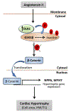
Fig. 6.. Proposed pathway for the role of OLA1 in ANG II-induced cardiac hypertrophy
Illustration of mechanism depicts that ANG II treatment increases OLA1 expression, which endogenously interacts with and phosphorylates GSK3β, rendering it inactive. Thus, β-catenin phosphorylation decreases (degradation reduces) leading to cellular accumulation and translocation to nucleus from cytosol resulting in transcription of cardiac hypertrophy response genes such as NPPA and MYH7. Sustained activation of these genes, which is a feature of cardiac hypertrophy, ultimately leads to heart failure. HW/TL- Heart Weight/Tibia Length.

References

    1. Ruilope LM, Schmieder RE: Left Ventricular Hypertrophy and Clinical Outcomes in Hypertensive Patients. Am J Hypertens 2008;21:500–508. - PubMed
    1. Sadoshima J, Izumo S: The Cellular and Molecular Response of Cardiac Myocytes to Mechanical Stress. Annu Rev Physiol 1997;59:551–571. - PubMed
    1. Frey N, Olson EN: Cardiac hypertrophy: the good, the bad, and the ugly. Annu Rev Physiol 2003;65:45–79. - PubMed
    1. Krishnamurthy P, Subramanian V, Singh M, Singh K: Beta1 integrins modulate beta-adrenergic receptor-stimulated cardiac myocyte apoptosis and myocardial remodeling. Hypertens Dallas Tex 1979 2007;49:865–872. - PubMed
    1. Aikawa R, Komuro I, Nagai R, Yazaki Y: Rho plays an important role in angiotensin II-induced hypertrophic responses in cardiac myocytes. Mol Cell Biochem 2000;212:177–182. - PubMed

Publication types

MeSH terms