Review
. 2019 May 6;49(3):375-391.
doi: 10.1016/j.devcel.2019.04.012.

Metastasis Organotropism: Redefining the Congenial Soil

Affiliations
Review

Metastasis Organotropism: Redefining the Congenial Soil

Yang Gao et al. Dev Cell. .

Abstract

Metastasis is the most devastating stage of cancer progression and causes the majority of cancer-related deaths. Clinical observations suggest that most cancers metastasize to specific organs, a process known as "organotropism." Elucidating the underlying mechanisms may help identify targets and treatment strategies to benefit patients. This review summarizes recent findings on tumor-intrinsic properties and their interaction with unique features of host organs, which together determine organ-specific metastatic behaviors. Emerging insights related to the roles of metabolic changes, the immune landscapes of target organs, and variation in epithelial-mesenchymal transitions open avenues for future studies of metastasis organotropism.

Keywords: EMT; immune microenvironment; metabolism; metastasis; niche; organotropism; seed and soil.


Figures

Figure 1.
Figure 1.. Bone-specific metastasis.
The bone microenvironment (ME) secretome generated by osteoblasts, osteoclasts or other cells may promote bone metastasis, while tumor cells can produce factors such as LOX to induce pre-metastatic niche formation. Interactions between tumor cells and osteoblasts through adherens junctions (via E-cadherin/N-cadherin, JAG1/Notch) and gap junctions (via CX43) also facilitate bone metastasis. Colonizing tumor cells express osteoblast-specific markers such as ALP and RUNX2 to escape immunosurveillance. In addition, tumor cells secrete factors promoting bone turnover to induce osteolysis, which in turn produces factors to stimulate tumor growth, creating a “vicious cycle”.
Figure 2.
Figure 2.. Liver-specific metastasis.
Hepatocytes directly interact with tumor cell enriched in claudin-2 to promote liver metastasis. They also promote the formation of a pro-metastatic niche though secretion of serum amyloid A1 and A2 (SAA). Hepatic stellate cells can produce PDGF, HGF, and TGFβ to induce liver metastasis. Tumor cells secrete exosomes, which are taken up by Kupffer cells. Integrin αvβ5 enriched exosomes stimulate Kupffer cells to produce pro-inflammatory S100A8, whereas MIF enriched exosomes trigger Kupffer cells to secrete TGFβ, which activates hepatic stellate cells, inducing liver-specific metastasis. LSECtin generated by sinusoidal endothelial cells also facilitates metastasis by inhibiting the T cell immune response.
Figure 3.
Figure 3.. Lung-specific metastasis.
Tumor-derived factors such as SPARC, VCAM1, and ANGPTL4, as well as EVs have been shown to be involved into tumor cell extravasation to the lung parenchyma. Tumor cells can activate TLR3 signaling in alveolar type II cells, which in turn recruit neutrophils and promote lung metastasis. Chemokines enriched in the lung such as CXCL12 and CCL21 recruit CXCR4 and CCR7 positive tumor cells. Alveolar macrophages can secrete the pro-inflammatory mediator Leukotriene B4 to suppress the T cell response and facilitate metastasis. Fibroblasts secrete CSTB to induce tumor cell survival. Also, fibronectin-enriched fibroblasts recruit VEGFR1 and integrin α4β1 positive hematopoietic progenitor cells to facilitate metastasis. Tumor cells also produce tenascin C to initiate metastasis and GALNT14 to overcome dormancy signals from fibroblasts. TSP-1 secreted from endothelial cells inhibits tumor cells self-renewal, while CX3CL1 expression leads to recruitment of CX3CR1-positive patrolling monocytes, preventing metastasis.
Figure 4.
Figure 4.. Brain-specific metastasis.
Tumor cells colonizing the brain need to produce cathepsin S, miR181c-enriched EVs and HPSE to overcome the defense provided by the blood-brain barrier. They also generate anti-plasminogen activator serpins to inhibit plasmin production from astrocytes to initiate metastasis. Astrocytes secrete many factors such as IL6, TGFβ, and IGF-1 that induce the growth of brain metastases. They also secrete miR19a-enriched exosomes, which inhibit PTEN expression and facilitate metastasis. Furthermore, gap junction characterized by CX43 between astrocytes and tumor cells and induced by PCDH7 stimulate brain metastasis. Exosomes enriched with miR503 induce M2 polarization of microglia which can promote metastasis through WNT signaling. Of note, bone and liver contain a fenestrated endothelium in contrast to the smooth lining of the lung endothelium and blood-brain barrier. Therefore, tumor cells develop different strategies to colonize at these different sites.
Figure 5.
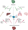
Figure 5.
Emerging facets of the “seed and soil” hypothesis. Green: organotropic cancer cells possess distinct metabolic features. Bone metastases utilize nutrients released during bone remodeling, such as serine, glycine, glucose, and glycerol. Liver metastases are highly glycolytic and consume local glucose. Lung metastases develop antioxidant strategies for survival in the pro-oxidant lung environment. Brain metastases exploit brain metabolites such as acetate, glutamine, amino acids and glutamine due to limited glucose resources. Blue: EMT regulates cancer organotropism. Liver metastases of pancreatic cancers require at least one copy of P120CTN, a stabilizer for membranous E-cadherin. Pancreatic cancer cells harboring homozygous P120CTN mutations can only metastasize to lung due to loss of E-cadherin. EMT may also contribute to organotropism through regulating metabolism and the immune microenvironment (ME). Red: different immune MEs regulates organ-specific metastasis. Bone metastasis is facilitated by osteoclasts, myeloid-derived suppressor cells (MDSCs), Tregs, and other bone-resident cells. Kupffer cells, CD11b/Gr1mid myeloid cells, metastasis-associated macarophages (MAMs), and neutrophils contribute to liver metastasis. Lung metastasis is regulated by alveolar macrophages, monocytes, MAMs, neutrophils, Tregs, and other cells. Microglia, the tissue-resident macrophages of the brain, promote brain metastasis.

References

    1. Aceto N, Bardia A, Miyamoto DT, Donaldson MC, Wittner BS, Spencer JA, Yu M, Pely A, Engstrom A, Zhu H, et al. (2014). Circulating tumor cell clusters are oligoclonal precursors of breast cancer metastasis. Cell 158, 1110–1122. - PMC - PubMed
    1. Aiello NM, Maddipati R, Norgard RJ, Balli D, Li J, Yuan S, Yamazoe T, Black T, Sahmoud A, Furth EE, et al. (2018). EMT Subtype Influences Epithelial Plasticity and Mode of Cell Migration. Dev Cell 45, 681–695.e4. - PMC - PubMed
    1. Albrengues J, Shields MA, Ng D, Park CG, Ambrico A, Poindexter ME, Upadhyay P, Uyeminami DL, Pommier A, Küttner V, et al. (2018). Neutrophil extracellular traps produced during inflammation awaken dormant cancer cells in mice. Science 361, eaao4227. - PMC - PubMed
    1. Altorki NK, Port JL, Mittal V, Markowitz GJ, McGraw T, Gao D, Stiles B, Saxena A, Port JL, Saxena A, et al. (2019). The lung microenvironment: an important regulator of tumour growth and metastasis. Nat Rev Cancer 19, 9–31. - PMC - PubMed
    1. Andrade K, Fornetti J, Zhao L, Miller SC, Randall RL, Anderson N, Waltz SE, McHale M, and Welm AL (2017). RON kinase: A target for treatment of cancer-induced bone destruction and osteoporosis. Sci Transl Med 9. - PMC - PubMed

Publication types

LinkOut - more resources