Review
. 2019 May 6;20(9):2217.
doi: 10.3390/ijms20092217.

Presynaptic Calcium Channels

Affiliations
Review

Presynaptic Calcium Channels

Sumiko Mochida. Int J Mol Sci. .

Abstract

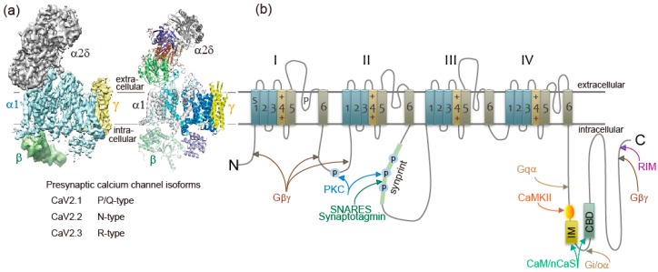
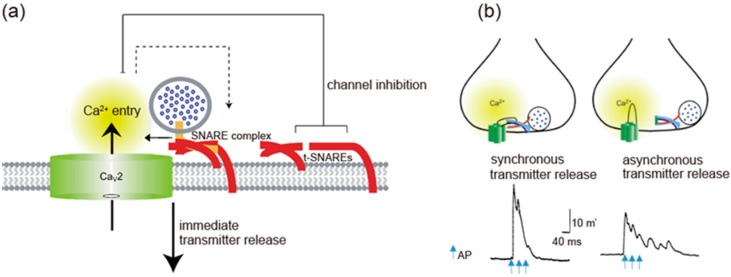
Presynaptic Ca2+ entry occurs through voltage-gated Ca2+ (CaV) channels which are activated by membrane depolarization. Depolarization accompanies neuronal firing and elevation of Ca2+ triggers neurotransmitter release from synaptic vesicles. For synchronization of efficient neurotransmitter release, synaptic vesicles are targeted by presynaptic Ca2+ channels forming a large signaling complex in the active zone. The presynaptic CaV2 channel gene family (comprising CaV2.1, CaV2.2, and CaV2.3 isoforms) encode the pore-forming α1 subunit. The cytoplasmic regions are responsible for channel modulation by interacting with regulatory proteins. This article overviews modulation of the activity of CaV2.1 and CaV2.2 channels in the control of synaptic strength and presynaptic plasticity.

Keywords: Ca2+ binding proteins; Ca2+ channels; G-proteins; synaptic proteins; synaptic transmission.


Conflict of interest statement

The authors declare no conflict of interest.

Figures

Figure 1
Figure 1
Ca2+ channel structure and organization. (a) The subunit composition and structure of high-voltage-activated Ca2+ channels. The cryo-EM structure of the rabbit voltage-gated Ca2+ channel Cav1.1 complex at a nominal resolution of 4.2 Å. The overall EM density map on the left is colored according to different subunits. The structure model on the right is color-coded for distinct subunits. Reproduced from [12]. (b) The α1 subunit consists of four homologous domains (I-IV), each consisting of six transmembrane segments (S1-S6). S1–S4 represents the voltage-sensing module. S5–S6 represents the pore-forming unit. The large intracellular loops linking the different domains of the α1 subunit serve as sites of interaction of different regulatory proteins important for channel regulation, including G-protein (Gβγ, Gα), RIM, SNARE proteins, and synaptotagmin at the synprint site (shown in green bar), calmodulin (CaM), and neuronal Ca2+ sensor proteins (nCaS) at the IQ-like motif, which begins with the sequence isoleucine-methionine (IM) instead of isoleucine-glutamine (IQ) and the nearby downstream CaM-binding domain (CBD), calmodulin kinase II (CaMKII), and protein kinase C (PKC). Adapted from [4].
Figure 2
Figure 2
Gβγ-mediated noradrenaline inhibition of transmitter release and N-terminal/I-II loop AID peptides of CaV2.2 α1-subunit. Noradrenaline (NA) induced Ba2+ current inhibition (upper traces) and transmitter release (lower graphs) were attenuated in the presence of Gβγ-interaction site of N-terminal peptide (CaV2.245-55, YKQSIAQRART) or AID peptide (CaV377-393, RQQQIEREL NGYLEWIF) (See Figure 1b). Ba2+ currents were recorded from superior cervical ganglion (SCG) neurons acutely dissociated from 3- to 6-week-old Wistar rats, while the synaptic transmission was recorded from long-term cultured SCG neurons isolated from p7 rat. NA was bath-applied 30 min after injection of the peptide at 1 mM in the injection pipette. EPSPs were normalized to amplitude prior to NA application at time = 0 min. Bar graph summarizing NA effects, *p < 0.05 vs. NA effects in controls (Student’s t-test). Adapted from [98].
Figure 3
Figure 3
Spatial regulation of transmitter release by the I-II loop interaction with SNAREs. (a) The I-II loop interacts with t-SNAREs, resulting in inhibition of Ca2.2 channels opening. Once AP opens the channels, an increase in Ca2+ mediates interaction with SNAREs complex and induces transmitter release. Adapted from [4]. (b) Triple APs induces a large synchronous transmitter release from the first AP. In contrast, asynchronous transmitter release was observed in the presence of 130 μM synprint peptide (see Figure 1b). Adapted from [99].
Figure 4
Figure 4
Temporal regulation of Ca2+ channel activity by CaM and nCaS after AP(s) firing modulates synaptic transmission. (a) Regulation of transmitter release (lower trace) after an AP firing (upper trace). Dependent on the inter-stimulus interval the second AP induces paired-pulse depression (PPD) and facilitation (PPF). The PPD was prevented by ΔCBD, while PPF was prevented by IM-AA mutation of CaV2.1 channels. (b) Model for Ca2+/CaM-dependent inactivation and facilitation of Ca2+ channels and neurotransmitter release. (c) Biphasic synaptic transmission during 1-s train of APs at 30 Hz changed to synaptic depression by the IM-AA mutation or to synaptic facilitation by the ΔCBD. (d) Overexpression of CaBP1 (blue) blocks synaptic facilitation, while overexpression of VILIP-2 (red) blocks synaptic depression, during 1-s train of APs at 10 Hz. Adapted from [90] (ac) and [113] (d).

References

    1. Dunlap K., Luebke J.I., Turner T.J. Exocytotic Ca2+ channels in mammalian central neurons. Trends Neurosci. 1995;18:89–98. doi: 10.1016/0166-2236(95)80030-6. - DOI - PubMed
    1. Snutch T.P., Reiner P.B. Ca2+ channels: Diversity of form and function. Curr. Opin. Neurobiol. 1992;2:247–253. doi: 10.1016/0959-4388(92)90111-W. - DOI - PubMed
    1. Tedford H.W., Zamponi G.W. Direct G protein modulation of Cav2 calcium channels. Pharm. Rev. 2006;58:837–862. doi: 10.1124/pr.58.4.11. - DOI - PubMed
    1. Catterall W.A., Few A.P. Calcium channel regulation and presynaptic plasticity. Neuron. 2008;59:882–901. doi: 10.1016/j.neuron.2008.09.005. - DOI - PubMed
    1. Tsien R.W., Lipscombe D., Madison D.V., Bley K.R., Fox A.P. Multiple types of neuronal calcium channels and their selective modulation. Trends Neurosci. 1988;11:431–438. doi: 10.1016/0166-2236(88)90194-4. - DOI - PubMed

MeSH terms

Substances

LinkOut - more resources